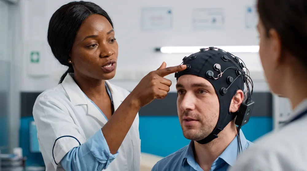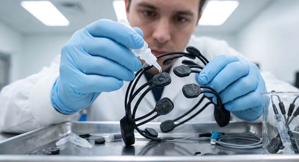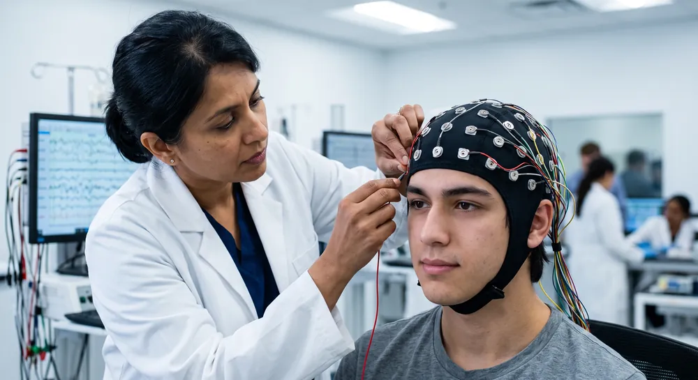
What separates a clear, interpretable neural signal from a trace corrupted by muscle artifact or line noise is not a sophisticated post-hoc algorithm, but a systematic, chronological pipeline of preparatory steps. EEG setup, in this context, is not simply the act of placing an electrode cap on a participant. It is a disciplined sequence of biological, environmental, and electrical calibration procedures that collectively establish a stable, low-noise interface between the brain and the amplifier.
This article walks through the pre-recording workflow, synthesizing available evidence to demonstrate that careful participant briefing, scalp preparation, environmental shielding, reference montage selection, and baseline artifact screening form a non-negotiable chain of dependencies.
What is an EEG Setup?
An EEG setup is the collection of equipment and procedures used to record electrical activity from the scalp. The term can refer to a clinical configuration for diagnostic evaluation, a research arrangement designed around a study protocol, or a portable system used outside a conventional laboratory. In each case, electrodes detect electrical potentials and an acquisition system turns them into a record that can be reviewed.
The setup also includes decisions about electrode locations, references, recording channels, and the conditions in which data are collected. Standardized placement supports consistency between recordings, while the chosen configuration should reflect the purpose of the examination or study.
EEG is used in clinical assessment and in neuroscience, among other areas. It records patterns of electrical activity rather than directly depicting brain structure, so findings are interpreted alongside the clinical or research context. An effective EEG setup therefore depends on both suitable equipment and careful recording practice.
Components of an EEG Setup
An EEG system brings together sensors, signal processing, and a means of recording the data. The components work as a chain: electrodes make contact with the scalp, the amplifier handles small electrical signals, and the recording system displays and stores the resulting channels. Fitting and connection details vary with the equipment and protocol.
EEG Electrodes
EEG electrodes detect electrical potentials at selected scalp locations. They may be placed individually or incorporated into a cap, with location determined by a standardized layout or a study-specific protocol. Contact quality matters because poor contact can contribute to unstable or noisy recordings; practical considerations are discussed in this overview of electrode contact quality.
Moreover, the selected electrode type and placement should match the recording needs, including the number and distribution of channels. Common setup considerations include:
Identifying the required scalp locations using the applicable placement system.
Preparing the contact area according to the electrode and equipment instructions.
Securing electrodes to reduce movement during the recording.
Checking connections and signal quality before proceeding.
These checks help distinguish equipment or contact problems from patterns that may be present in the recording.
EEG Amplifier
The amplifier receives the small voltage differences detected by the electrodes and makes them available for recording and display. Its channel capacity and compatible inputs should align with the planned electrode arrangement. The amplifier is one part of the signal chain, so reliable connections and appropriate system configuration matter as much as the device itself.
EEG Machine Set Up and Recording System
The recording system connects to the amplifier and presents EEG channels for observation, storage, and later review. Before data collection, the operator configures the recording according to the protocol, confirms that channels are available, and checks that the display is behaving as expected. The exact controls and workflow depend on the system in use.
The roles of common system elements can be summarized without assuming that every setup uses identical hardware or software.
Element | General role | Setup consideration |
|---|---|---|
Electrode inputs | Receive signals from scalp electrodes | Match connections to the intended channel map |
Amplifier | Handles the small electrical signals | Confirm compatible inputs and stable connections |
Recording interface | Displays and stores channels | Check configuration and data capture |
Supporting equipment | Helps maintain the recording arrangement | Follow applicable equipment and site procedures |
A functioning display alone does not establish that a recording is technically sound. Channel checks, observation of signal behavior, and documentation of the configuration help make the collected data easier to review and interpret.
EEG Cap or Headband
An EEG cap or headband holds electrodes in a repeatable arrangement and can simplify placement when multiple sites are needed. Fit, electrode spacing, and compatibility with the recording protocol affect how well a particular cap serves the task. A cap does not replace the need to verify the location and contact of its electrodes.
Types of EEG Setups
EEG setups are often grouped by where and why recording takes place. The distinction is useful, but the categories can overlap: a research study may use clinical equipment, and a portable system may collect data for a clinical or research purpose. Each arrangement must still support clear recording and appropriate documentation.
Clinical EEG Setup
A clinical EEG setup is arranged for recording that contributes to a medical evaluation. The electrode plan, recording duration, and any procedures during the examination are determined by the clinical question and local protocol. The resulting EEG is reviewed by qualified professionals in context; the recording itself is not a standalone diagnosis.
Research EEG Setup
A research EEG setup follows the study design, including its required electrode locations, recording conditions, and data-management plan. Consistent procedures across participants and sessions support comparison, while metadata can help document differences that may affect interpretation. Research protocols also address consent, privacy, and the handling of recorded data.
Portable EEG Setup
A portable EEG setup is designed to be used away from a fixed laboratory or examination room. Portability can make recording possible in a broader range of environments, although movement, electrical interference, and changes in recording conditions may introduce practical challenges. The system and protocol need to account for the setting in which data are collected.
Why Participant Briefing is Crucial for Reliable EEG Data
Briefing and protocol standardization are designed to reduce procedural variance that arises from unpredictable behavior.
A participant who understands the task instructions and the importance of minimizing facial tension, swallowing, and blinking will produce fewer high-amplitude muscle artifacts that can confound posterior signal analysis. The process of obtaining consent for skin preparation steps also establishes a collaborative dynamic, preempting surprise movements during scalp cleaning.
An extensive review on managing EEG studies emphasizes that developing formal written protocols, training teams, and conducting pilot sessions are essential to ensuring consistent implementation across multiple recording sessions and sites. The review, which draws on large-scale multi-site experience, explicitly ties pre-data-collection planning to the validity of the final dataset. Without this foundational standardization, even the most advanced EEG headset cannot compensate for the noise introduced by inconsistent participant state.
4 recommendations from the review:
Establish formal written protocols and team training for multi-session consistency
Run pilot sessions to identify setup issues before real data collection
Standardize participant instructions around movement, jaw clenching, and blinking
Treat behavioral standardization as a critical upstream noise control
Why Preparation Time Directly Impacts EEG Recording Fidelity
Once the participant is briefed, the workflow shifts to the biological interface. The scalp must be prepared to create a low-impedance, stable connection between the skin and the recording electrodes.
Typical preparation involves cleaning the scalp with an alcohol solution and, for wet electrode systems, lightly abrading the skin to remove dead cells and oils. This step is often time-consuming, and there is a natural desire to accelerate it, especially in clinical or consumer-facing contexts. However, a direct evaluation of this trade-off comes from a 2024 BCI study that compared fast-setup systems using dry and wet electrodes.
In that study, the average system setup time for dry electrodes was 38.4 seconds, while the wet electrode procedure took 103.4 seconds. Signal quality, as measured by the information transfer rate in a steady-state visual evoked potential (SSVEP) paradigm, differed meaningfully: the wet setup achieved 138.89 bits per minute, compared to 70.59 bits per minute for the dry setup.
Although both configurations produced functional BCI performance, the faster dry setup introduced a higher noise floor. The direct correlation between preparation time and recording fidelity is explicit: a slower, more thorough biological interface preparation yielded a cleaner signal.
The exact shape of this time-to-quality curve is context-dependent and cannot be generalized across all electrode types and scalp conditions, but the BCI data provides the clearest available evidence that the investment in scalp preparation is not optional, it directly determines the raw data quality. A fast EEG machine setup may be marketed as a convenience, but it inherently compromises the biological interface unless alternative electrode technologies can close the gap.
Metric | Dry setup | Wet setup |
|---|---|---|
Setup time | 38.4 seconds | 103.4 seconds |
Information transfer rate | 70.59 bits/min | 138.89 bits/min |
Noise floor | Higher | Lower |
Best Practices for Mitigating Environmental Noise in EEG Labs
With the participant prepared, the next step is to control the physical recording environment. Electrical noise, particularly 50 or 60 Hz line noise from power mains, can easily infiltrate a high-gain amplifier if the recording space is not properly arranged.
Therefore, the standard workflow involves positioning the participant away from unshielded power sources, turning off non-essential electrical devices, and using grounded, shielded enclosures where possible. Ambient temperature and lighting also matter since fluctuations can induce sweat artifacts or photic stimulation that alters ongoing brain rhythms.
The aforementioned EEG study management review explicitly recommends planning the physical environment and equipment setup during the pilot phase. By testing the recording conditions in advance, teams can identify and eliminate sources of external interference before formal data collection begins.
While the review does not provide quantitative benchmarks for shielding effectiveness, such as a decibel reduction from a Faraday cage, the procedural principle is consistent: a standardized environment reduces extrinsic variance that would otherwise masquerade as neural activity. For portable EEG applications, where a fully shielded room is often impractical, the same principle applies in a different form: the operator must systematically document and, where possible, eliminate nearby noise sources during the pre-recording checklist.
The absence of direct quantitative evidence in the provided studies means that specific shielding claims remain inferred from engineering consensus rather than empirically validated in the EEG literature, but the logical necessity of environmental control is not in dispute.
Choosing a Referencing Strategy During EEG Setup
A less visible but equally consequential decision in the pre-recording workflow is the choice of reference montage. Every voltage measurement in EEG is a difference between two points, and the reference electrode defines what is treated as “zero” for all other channels.
The decision of where to place the reference and how to mathematically re-reference the data is often deferred to the analysis stage, but the physical montage and the conceptual referencing strategy are setup-phase choices that fundamentally shape the spatial topography of the recorded signal.
A comparative pre-processing pipeline study evaluated several common re-referencing approaches: Common Average Reference (CAR), robust CAR (rCAR), Reference Electrode Standardization Technique (REST), and its interpolated variant (RESIT). The analysis revealed that while CAR, REST, and RESIT produced similar topographical representations, the rCAR method generated notably different event-related spectral perturbation (ERSP) patterns. Signal segmentation also significantly affected the cleaning outcome.
These findings confirm that the choice of referencing scheme is a parameter-dependent methodological step that biases the data from the moment of acquisition. During the setup, the placement of the reference electrode (often on the earlobe or mastoid) and the planned re-referencing logic must therefore be decided and documented.
A casual or unconsidered reference selection can later produce misleading cortical maps, making this a substantive rather than a trivial aspect of the EEG cap and system configuration.
How to Screen for Artifacts During EEG Baseline Calibration
After the participant is positioned, the scalp is prepared, the environment is shielded, and the reference is chosen, the final gateway before recording is a baseline calibration period. This typically involves a short resting-state segment, often with eyes open or eyes closed, during which the experimenter verifies that the amplifier is stable, the electrode-skin interfaces are balanced, and the signal is free from obvious artifacts such as excessive DC drift, high-frequency muscle noise, or slow ocular potentials.
The calibration period serves as a diagnostic snapshot: a loose electrode will show a wandering baseline, a poorly abraded site will display elevated impedance and line noise, and a tense participant will generate persistent muscle activity.
The EEG study management review stresses the importance of ongoing data quality monitoring and using pilot sessions to catch setup-related issues early. Meanwhile, the pre-processing pipeline comparison found that how the continuous signal is segmented into epochs significantly affects the subsequent cleaning procedure.
This has direct implications for baseline calibration since the operator must decide what constitutes an acceptable “clean” rest epoch, and that decision will influence artifact rejection thresholds applied later in the analysis. For consumer-grade home EEG devices, automated impedance checks and built-in artifact indicators partially codify this calibration, but the fundamental logic is the same: a deliberate pause to screen the raw signal before data collection prevents the storage of epochs that will eventually be rejected during pre-processing.
The Impact of Fast EEG Setups on Data Rigor
The pre-recording EEG workflow is a sequence of interdependent steps, and each step represents a decision point where time can be saved at the expense of signal quality.
The BCI fast-setup study provides the most direct illustration of this trade-off. A dry electrode application took just 38.4 seconds, which is less than one-third the time required for the wet electrode setup, but the resulting signal quality was measurably lower. Task performance, in that specific BCI context, was still adequate, but the elevation of the noise floor is an inescapable consequence of an abbreviated biological interface preparation. Skipping or shortening the scalp cleaning, the environmental checks, or the baseline screening will not necessarily render the recording unusable, but it will invariably reduce the clean signal bandwidth available for subsequent analysis.
It is important to acknowledge that the direct evidence for many of these individual workflow steps is limited. The study management review supports the value of protocol standardization, environmental planning, and pilot screening, but it does not isolate the causal contribution of, say, a specific briefing script or a particular shielding distance on final signal-to-noise ratio.
The montage selection study shows that re-referencing choices affect outcomes, but it evaluates post-acquisition processing rather than the raw data quality at the physical setup stage. The BCI study offers a concrete duration-to-quality comparison, but its generalizability to other electrode systems and recording contexts is unproven.
Consequently, the recommendations outlined here are best understood as a framework for systematic practice, derived from the best available evidence and the consensus of the field, rather than as empirically proven universal mandates. The central message is that careful pre-recording calibration is the single most critical factor in obtaining clean, interpretable neural signals, and that every shortcut taken during the setup phase is a direct trade against the fidelity of the final dataset.
How EEG Setup Differs Across Emotiv Systems
The pre-recording workflow applies to every EEG system, but how long each step takes, and where its risks sit, depends largely on the sensor interface. Emotiv's wireless EEG systems are designed to take research-grade recording beyond the fixed laboratory and into classrooms and mobile field studies. Because the range covers gel, saline, semi-dry and dry sensors, a research team can choose where to sit on the preparation-time and signal-quality trade-off described above, rather than building every protocol around a single setup.
System | Channels | Sensor interface | What preparation involves |
|---|---|---|---|
Up to 32 | Sintered Ag/AgCl sensors used with gel | Conventional wet preparation, with sensors placed in a cap with 74 openings at standard 10-20 positions | |
Up to 32 | Refillable saline felt sensors | Felt pads are wetted, and a rear refill opening keeps conductivity consistent without interrupting the study | |
14 | Saline-soaked felt pads | Pads are re-wetted before each session and can dry out during extended recordings | |
5 | Semi-dry polymer sensors | A few drops of solution; setup takes about 1–2 minutes | |
16 | Interchangeable dry flat and comb sensors (saline felt pads also fit) | Nothing to prepare and no cleanup between participants | |
2 | Dry in-ear elastomer sensors | Setup in under a minute |
Narrowing the Dry-Electrode Gap
The BCI study discussed above found that a fast dry-electrode setup came with a higher noise floor, a trade-off that holds unless alternative electrode technologies can close the gap. EPOC X PRO addresses this at the sensor itself. Each sensor has its own active amplifier, which reduces interference from cable movement and electrode impedance, and the signal is acquired at 24-bit resolution.
The extra headroom lets low-frequency drift, electrode offset and sudden movement artifacts be captured without clipping, leaving fewer trials to reject during pre-processing. Emotiv designed this combination so that dry sensors can deliver signal quality comparable to wet sensors.
Sensor geometry matters as well. Flat sensors sit on bare skin at the forehead and mastoid reference sites, while comb sensors, available in two lengths, reach through hair. Any sensor type clips into any socket, so researchers can match the sensor to each site instead of compromising with one geometry across the whole scalp. And because dry sensors do not rely on saline, session length is not limited by how long a pad stays wet, which matters for extended workload studies and protocols with many participants.
None of this replaces the baseline check. A short resting segment remains the way to confirm signal quality for a given protocol and participant group.
Carrying the Workflow Out of the Lab
Each step of the pre-recording workflow still applies outside a shielded room; what changes is how it is carried out.
Environment: Emotiv systems are wireless, so no amplifier cable tethers the participant to a workstation. Epoc X and Flex 2 use common-mode noise rejection, and EPOC X PRO adds Wi-Fi 6, which holds up better in the busy 2.4 GHz conditions common in real-world research settings.
The 9-axis motion sensors in Epoc X, Insight, Flex 2 and EPOC X PRO record head movement alongside the EEG, so movement during a session is documented rather than guessed. EPOC X PRO can also record on board for more than 8 hours at 256 Hz without a connected computer, so a recording continues if the wireless connection drops.
Reference: Reference placement is part of each system's design, which makes this setup-phase decision straightforward to document. Epoc X places its CMS/DRL references at P3/P4, with the mastoids as an alternative. EPOC X PRO fixes CMS on the left mastoid and DRL on the right, which frees P3 and P4 to record EEG.
Insight references on the left mastoid, MN8 uses split CMS/DRL sensors on the back of the ear, and Flex 2 lets researchers place CMS/DRL at any 10-20 location or on the ears.
Baseline check: Epoc X, Insight, Flex 2 and MN8 include a real-time contact quality monitor. EPOC X PRO builds a contact quality indicator light into each sensor housing, so the operator can check fit at the sensor itself, even when no host device is connected.
In EmotivPRO, event markers can label eyes-open and eyes-closed baseline segments, and Lab Streaming Layer (included in the Standard Team and Performance plans) synchronizes EEG with stimulus software such as PsychoPy.
Matching the System to the Protocol
For high-density studies that need flexible electrode placement, Flex 2 Gel or Flex 2 Saline brings up to 32 channels into a wireless cap. Epoc X suits academic research, BCI development and classroom work where 14 channels are enough and sessions are short enough that saline preparation is not a burden.
EPOC X PRO is built for long sessions, field studies and timing-sensitive research, with timestamps synchronized to within about 1 millisecond. Insight supports portable BCI development and demonstrations, and MN8 brings EEG into an earbud for everyday use with the Emotiv App.
Because Epoc X and EPOC X PRO share 14 core recording sites and the same EmotivPRO and Cortex software, teams can also combine them, for example piloting and training on Epoc X while running core recordings on EPOC X PRO. The hardware comparison sets the specifications side by side.
Why Careful EEG Preparation Decides the Value of Every EEG Recording
The central lesson from the entire pre-recording workflow is that the quality of a neural signal is fixed before data collection begins, not rescued afterward by clever analysis. Each stage—from briefing the participant to cleaning the scalp, shielding the room, choosing the reference, and checking the baseline—forms a chain where a weak link sends noise into every later measurement.
The trade-off translates to shorter setup saves time but raises the noise floor, as shown by the direct comparison between fast dry electrodes and slower wet electrodes. No post-processing filter can fully separate the brain's true electrical activity from artifacts introduced by movement, environmental interference, or a poorly prepared sensor interface.
Given that direct quantitative evidence for many individual steps remains limited, the workflow is best treated as a disciplined framework rather than a set of proven mandates. The strongest available data points to preparation time as a measurable factor in signal fidelity, while other recommendations rest on field consensus and engineering logic.
What is beyond dispute is that a deliberate, systematic pause before recording prevents wasted sessions and misleading cortical maps. In short, the most expensive and powerful EEG system is only as trustworthy as the careful calibration that precedes it.
References
Boudewyn, M. A., Erickson, M. A., Winsler, K., Ragland, J. D., Yonelinas, A., Frank, M., ... & Luck, S. J. (2023). Managing EEG studies: How to prepare and what to do once data collection has begun. Psychophysiology, 60(11), e14365. https\://doi.org/10.1111/psyp.14365
Li, X., Wang, J., Cao, X., Huang, Y., Huang, W., Wan, F., ... & Xie, S. Q. (2024). Evaluation of an online SSVEP-BCI with fast system setup. Journal of Neurorestoratology, 12(2), 100122. https\://doi.org/10.1016/j.jnrt.2024.100122
Coelli, S., Calcagno, A., Cassani, C. M., Temporiti, F., Reali, P., Gatti, R., ... & Bianchi, A. M. (2024). Selecting methods for a modular EEG pre-processing pipeline: An objective comparison. Biomedical Signal Processing and Control, 90, 105830. https\://doi.org/10.1016/j.bspc.2023.105830
Frequently Asked Questions
Why is participant briefing considered the first step in ensuring good EEG signal quality?
Briefing reduces procedural variance by teaching participants to minimize facial tension, swallowing, and blinking, which are major sources of muscle artifact. It also establishes consent and collaboration, preventing surprise movements during scalp preparation. This behavioral standardization acts as the first biological noise filter before any physical setup begins.
How does scalp preparation time affect the quality of an EEG recording?
Longer, more thorough scalp preparation creates a low-impedance, stable connection between the skin and electrodes, which lowers the noise floor. Evidence from a brain-computer interface study showed that a slower wet-electrode setup produced a cleaner signal than a faster dry-electrode setup. The investment in preparation time directly determines the raw data quality.
What are the key environmental controls needed before recording an EEG?
The participant should be positioned away from unshielded power sources, non-essential electrical devices should be turned off, and grounded or shielded enclosures should be used when possible. Ambient temperature and lighting must also be controlled because fluctuations can cause sweat artifacts or alter brain rhythms. A standardized environment reduces extrinsic electrical noise that could otherwise masquerade as neural activity.
Why is the choice of reference montage an important part of EEG setup?
Every EEG voltage measurement is a difference between two points, so the reference electrode defines what is treated as "zero" for all other channels. Different referencing strategies, such as Common Average Reference or REST, can produce different topographic and spectral results. A casual or unconsidered reference selection can later generate misleading cortical maps, making this a substantive setup-phase decision.
What is the purpose of the baseline calibration period before recording?
Baseline calibration provides a short resting-state segment to verify that the amplifier is stable, electrode-skin interfaces are balanced, and the signal is free from obvious artifacts. It acts as a diagnostic snapshot to catch loose electrodes, poor scalp sites, or muscle tension before data collection begins. This deliberate screening prevents the storage of epochs that would later be rejected.
What is the single most critical factor in obtaining clean, interpretable neural signals?
Careful pre-recording calibration is the single most critical factor, as it establishes a stable, low-noise interface between the brain and the amplifier. The quality of an EEG recording is largely determined before the first data epoch is stored, not by post-hoc algorithms. A weakened step in the preparatory chain propagates noise downstream and degrades the final signal.
Emotiv is a neurotechnology leader helping advance neuroscience research through accessible EEG and brain data tools.
Medical Disclaimer: The information provided on this website is for educational and informational purposes only and is not intended as medical or health advice. This content may contain errors and should not be relied upon to make life-altering health, medical, or lifestyle choices. Always seek the advice of your physician or other qualified health provider with any questions you may have regarding a medical condition or treatment.
Christian Burgos