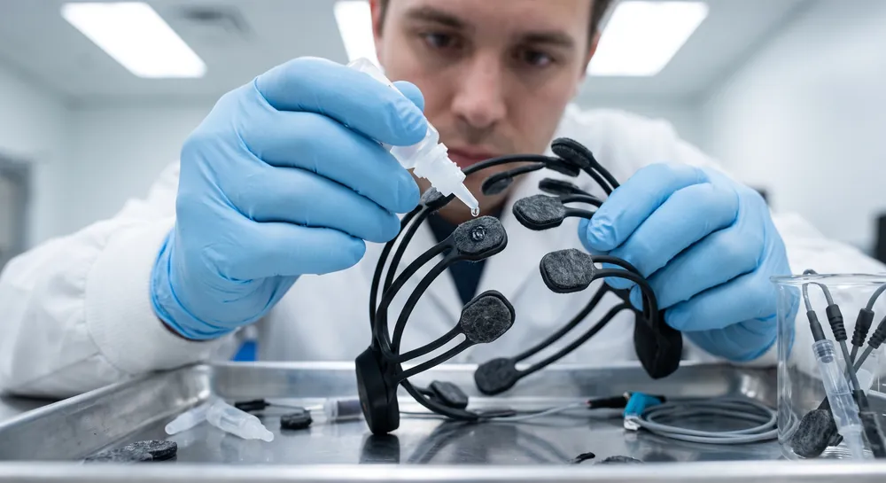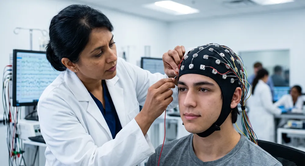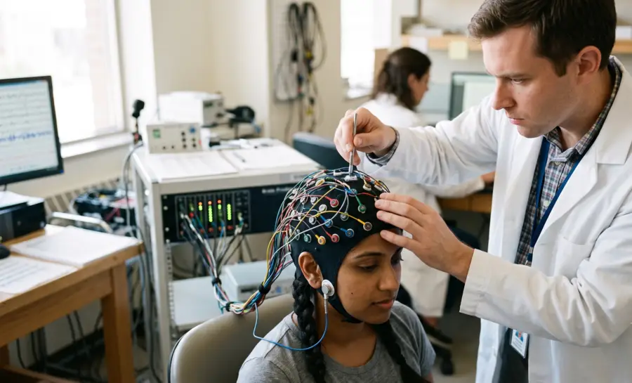
A referential montage takes the voltage recorded at each active electrode on the scalp and subtracts it from the voltage recorded at a single, shared reference point.
The math is simple. The consequences are not.
This single subtraction step determines the shape, size, and apparent location of every wave that ends up on the page, and the electroencephalogram itself is only as trustworthy as the reference behind it.
What is an EEG Reference Electrode?
The Role of the Reference Electrode in EEG
Every electrical measurement requires a stable point of comparison, and in EEG studies, this is the function of the reference electrode. Because the hardware measures voltage differences between two distinct points on the scalp, the reference provides the relative "zero" for the electrical activity detected by other sensors.
Without this baseline, it would not be possible to isolate the neural rhythms that represent complex neuroscience phenomena. By subtracting the signal recorded at the reference from the signal at an active electrode, the amplifier produces a clean output that reflects neural activity excluding common-mode noise.
EEG Reference Electrode Placement: Types Explained
Selecting a location for the reference electrode influences the interpretation of the brain's spatial activity.
Common sites include the mastoid bones or the earlobes, which are selected because they are relatively inactive zones compared to the regions over the cerebral cortex. Researchers sometimes utilize a linked-ear configuration to mitigate individual differences.
Certain high-density arrays allow for average reference, where researchers calculate the mathematical mean of all electrodes to serve as a virtual reference point, minimizing bias introduced by a single inferior location.
Why Reference Choice Matters: The Core Problem
The logic of a referential recording can be written as a simple subtraction:
Signal \= Active Electrode – Reference Site
Change the reference, and you change the subtraction. That means amplitude shifts, wave shapes distort, and the apparent scalp location of an event can move without anything in the brain having changed at all.
There is a common assumption in clinical and research settings that a carefully chosen reference will reveal the “true” local activity at each electrode, stripped of contamination. This assumption has intuitive appeal, but it has only been tested rigorously in a narrow set of clinical contexts.
The studies that have tested it directly show a more complicated picture, one where the referential montage sometimes performs well and sometimes actively misleads the interpreter about where activity in the brain is actually happening.
How Referential Montages Misplace Brain Activity
The clearest demonstration of this problem comes from research on corticocortical evoked potentials, or CCEPs. These are electrical responses generated when a small pulse of stimulation is delivered to one part of the brain and a response is recorded at another site, a technique used to map how different brain regions communicate.
Researchers led by Dickey et al., using depth electrodes (thin probes inserted directly into brain tissue) compared how well a referential montage could correctly identify whether a given electrode contact was sitting in gray matter (where neuron cell bodies cluster and most functional processing happens) or white matter (the connective wiring between regions, which generates far less of its own electrical activity).
The results were stark. Using a referential montage, only 12 of 27 electrode contacts, or 44%, showed a significantly higher amplitude when positioned in gray matter compared with white matter.
A Laplacian montage, which calculates activity at each electrode relative to the average of its immediate neighbors rather than a single distant reference, correctly identified 25 of 27 contacts, or 93% (P \= 0.0003). When researchers measured how reliably each EEG montage could classify a contact as gray or white matter using a statistical measure called area under the curve (a score of 1.0 means perfect classification, while 0.5 means no better than a coin flip), the referential montage scored 0.51, essentially chance performance.
The signals were frequently and falsely pointing to white matter as the source of activity that was actually generated elsewhere.
Moreover, a second study by Otero et al. reinforces how much reference choice can shift the apparent findings, even when a real underlying difference between groups exists. Researchers comparing iron-deficient schoolchildren to iron-replete peers analyzed the same underlying EEG data using two different montages.
The referential montage highlighted excess delta activity (a slow brain wave frequency) concentrated in the frontal regions of iron-deficient children. The Laplacian montage, applied to the identical dataset, instead revealed widespread excess theta activity (a slightly faster slow-wave frequency) spread across the whole scalp.
The children were the same. The recording sessions were the same. The only variable was the montage, and it changed both the frequency band flagged as abnormal and the region of the brain where that abnormality appeared to live.
Together, these two studies establish a working principle: a referential montage can genuinely mislead localization, and even when a real difference between groups exists in the underlying data, the topography of where that difference appears to be located is shaped heavily by which montage was used to look at it.
Study | Comparison | Key Result |
|---|---|---|
CCEP gray/white | Referential vs Laplacian | Referential mislocalized to white matter |
Iron deficiency children | Referential vs Laplacian | Montage shifted abnormal frequency, region |
Ipsilateral vs. Contralateral Ear Reference: Which Works Better
If the reference site itself is a variable, does it matter which ear you pick when using an ear-referenced montage?
A study by Bubrick et al. assessing a simplified “hairline” EEG setup, a reduced electrode arrangement designed for rapid bedside screening, tested this directly in the context of detecting nonconvulsive status epilepticus, a state of ongoing seizure activity without the visible convulsions typically associated with seizures.
Researchers reformatted standard EEG recordings into three abbreviated montages:
A bipolar montage (comparing pairs of adjacent electrodes to each other rather than to a distant reference)
A referential montage to the ear on the same side as each active electrode (ipsilateral)
Referential montage to the ear on the opposite side (contralateral)
Five neurophysiologists then interpreted the reformatted samples and their readings were compared against the original full-montage interpretation.
Bipolar montage: 71% correct interpretations
Ipsilateral ear reference: 70.5% correct
Contralateral ear reference: 65% correct
This gap suggests that referencing to the ear on the same side as the electrode being measured preserves more diagnostic accuracy than referencing across the head to the opposite ear.
But the more important finding sits beneath that comparison. Even with the best-performing montage, sensitivity for detecting actual seizures was only 72%, and seizures were frequently misread as more benign patterns, including normal recordings or diffuse slowing.
The takeaway is not simply that ipsilateral referencing is the better technical choice. It is that even the best version of this stripped-down referential setup missed more than a quarter of seizures, which makes it insufficiently reliable as a tool to rule out nonconvulsive status epilepticus in a patient where the stakes of a missed diagnosis are high.
Cz Reference in the ICU: A Pragmatic Success
Not every referential setup performs poorly. A separate 2010 study designed a seven-electrode montage (Fp1, Fp2, T3, T4, O1, O2, and Cz) specifically for rapid seizure screening in critically ill patients, using the vertex electrode Cz as the shared reference point for all channels.
The appeal of this design was practical: it can be applied using only anatomic landmarks such as the pupils, ears, vertex, and inion, without a tape measure, and it can be placed and interpreted quickly by residents when full technical EEG support is not available.
When full 10-20 system recordings from critically ill patients were reformatted into this simplified Cz-referential montage and reviewed independently by neurology attendings and senior residents, the average sensitivity for seizure detection was 92.5%, with a specificity of 93.5%. These numbers stand in sharp contrast to the 72% sensitivity found in the ear-referenced hairline montage study above, suggesting that the choice of Cz as a reference, combined with this particular seven-electrode layout, may capture seizure activity more reliably than an ear-based alternative in this setting.
That said, the study was retrospective and drew from a small sample, and the authors themselves state plainly that prospective validation in a larger population is still needed before this can be treated as an established clinical tool.
When Referential Montages Add Unique Localizing Value
The picture shifts again in a different clinical scenario. Localizing seizures originating from the mesial temporal lobe, a deep brain structure associated with memory and frequently implicated in epilepsy.
Researchers led by Parcia SV reviewed 76 ictal (seizure-time) recordings using both sphenoidal electrodes, thin electrodes placed near the base of the skull close to the temporal lobe, and standard scalp electrodes, analyzing the data in both bipolar and referential montages.
In patients with mesial temporal lobe epilepsy, seven of the seizures recorded from three patients showed activity confined exclusively to a single sphenoidal electrode before any scalp electrode showed involvement, and this pattern was visible using the referential montage. The bipolar montage did not reveal this same exclusive early activity.
This isolated early pattern occurred only in patients with mesial temporal lobe epilepsy and did not appear in neocortical temporal lobe epilepsy, where sphenoidal and scalp electrodes showed simultaneous involvement regardless of which montage was used (p \< 0.04 for the association between this early sphenoidal-only pattern and mesial onset).
This is a meaningful counterpoint to the localization failures described earlier. In this specific clinical context, deep-source seizure activity near the sphenoidal electrode, a referential montage picked up an early localizing signal that a bipolar montage missed.
The benefit appears tied closely to this particular anatomical scenario rather than serving as a general rule that referential montages outperform other approaches.
Recognizing Reference-Related Artifacts
Because every channel in a referential recording is calculated against the same single point, any noise contaminating that reference electrode gets distributed across the entire recording. A muscle twitch, an eye movement, or a poorly seated electrode at the reference site does not just corrupt one channel. It appears, inverted, in every channel simultaneously.
A practical example: if a mastoid reference electrode is picking up muscle activity from jaw clenching, that rhythmic muscle signal will be superimposed on every channel in the montage, potentially mimicking a widespread rhythmic pattern that looks like it originates in the brain itself when it is actually an artifact of the reference site.
This raises an unresolved question about the iron deficiency study discussed earlier. The frontal delta excess detected using the referential montage sits in a region of the scalp close to the eyes, where eye-movement artifact commonly contaminates recordings.
The study did not test whether eye movement contributed to this finding, and no conclusion should be drawn that it did. But the possibility illustrates why any topographic finding produced by a referential montage, particularly one localized to frontal regions, deserves a second look before being accepted as a genuine cerebral pattern rather than a reference-site artifact.
4 Ways to Mitigate Reference-Related Pitfalls
A few practical habits reduce the risk of being misled by reference-related distortion.
Always identify what the reference electrode is before interpreting a tracing. If an identical or near-identical waveform shows up simultaneously across every channel, that pattern points toward a reference artifact rather than a genuine, widespread brain signal.
Cross-check findings with a different montage whenever possible. The CCEP localization study and the iron deficiency study both demonstrate that Laplacian or bipolar montages can correct false gray-matter localizations and clarify which frequency band and scalp region are truly involved, rescuing interpretations that a referential montage alone would have distorted.
When using a simplified referential setup for rapid screening, such as a hairline montage or a seven-electrode ICU configuration, compare its performance against a full, gold-standard recording before trusting it in a high-stakes decision. This is precisely the comparison performed in the ICU seizure detection study and the critique applied to the hairline screening study.
For presurgical evaluation and other high-stakes localization tasks, do not rely on a referential montage in isolation. Combine it with other montages and clinical context, following the approach used in both the CCEP localization work and the sphenoidal electrode study on mesial temporal lobe epilepsy.
Summary
A referential montage is straightforward to set up and, in selected circumstances, can provide clinically useful information that other montages miss, as seen in the Cz-referenced ICU seizure screening and in the early sphenoidal localization of mesial temporal lobe seizures. But its output is shaped profoundly by the reference site chosen, and that dependency can produce false localizations, as seen in the depth-electrode CCEP research, or miss a substantial share of seizures outright, as seen in the hairline screening comparison of ear references.
Many of the reference choices used routinely in clinical and research settings, including linked ears and mastoid processes, have not been subjected to the kind of head-to-head comparison seen in these studies. Their reliability is often assumed rather than demonstrated. This gap matters for anyone working with neuroscience data drawn from EEG, whether in a hospital, a research lab, or a classroom studying brain signals for the first time.
The most useful habit when reading any referential EEG tracing is to ask two questions before interpreting a single wave: what is the reference electrode, and what activity, cerebral or otherwise, might it be contributing to every channel on the page?
References
Dickey, A. S., Alwaki, A., Kheder, A., Willie, J. T., Drane, D. L., & Pedersen, N. P. (2022). The Referential Montage Inadequately Localizes Corticocortical Evoked Potentials in Stereoelectroencephalography. Journal of clinical neurophysiology : official publication of the American Electroencephalographic Society, 39(5), 412–418. https://doi.org/10.1097/WNP.0000000000000792
Otero, G. A., Aguirre, D. M., Porcayo, R., & Fernández, T. (1999). Psychological and electroencephalographic study in school children with iron deficiency. The International journal of neuroscience, 99(1-4), 113–121. https://doi.org/10.3109/00207459908994318
Bubrick, E. J., Dworetzky, B. A., & Bromfield, E. B. (2007). Assessment of hairline EEG as a screening tool for nonconvulsive status epilepticus. Epilepsia, 48(12), 2374–2375. https://doi.org/10.1111/j.1528-1167.2007.01260_4.x
Karakis, I., Montouris, G. D., Otis, J. A., Douglass, L. M., Jonas, R., Velez-Ruiz, N., ... & Espinosa, P. S. (2010). A quick and reliable EEG montage for the detection of seizures in the critical care setting. Journal of Clinical Neurophysiology, 27(2), 100-105. https://doi.org/10.1097/wnp.0b013e3181d649e4
Pacia, S. V., Jung, W. J., & Devinsky, O. (1998). Localization of mesial temporal lobe seizures with sphenoidal electrodes. Journal of clinical neurophysiology, 15(3), 256-261. https://doi.org/10.1097/00004691-199805000-00010
Frequently Asked Questions
What is a referential EEG montage?
A referential montage subtracts the voltage at a single shared reference electrode from the voltage at each active scalp electrode. This single subtraction shapes the amplitude, wave shape, and apparent location of every brain signal displayed.
Why does changing the reference electrode alter what the EEG shows?
The displayed signal equals the brain activity under the active electrode minus whatever activity is present at the reference site. Choosing a different reference changes that subtraction, which can shift amplitudes, distort wave shapes, and move the apparent source of an event.
Can a referential montage mislead about where brain activity originates?
Yes. In depth-electrode studies, a referential montage performed no better than chance at distinguishing gray matter from white matter activity, while a Laplacian montage correctly identified the vast majority. Another study found that the referential and Laplacian montages flagged different frequency bands and different scalp regions for the same dataset, showing the montage heavily shapes topography.
Which ear reference is more reliable, ipsilateral or contralateral?
In a hairline EEG setup for detecting nonconvulsive seizures, referencing to the ipsilateral ear (same side) produced slightly higher diagnostic accuracy than referencing to the contralateral ear. However, even the better ipsilateral configuration missed a substantial share of seizures, making it insufficient for ruling out the condition.
How did a Cz-referenced montage perform in ICU seizure screening?
When Cz was used as the reference in a simplified seven-electrode layout, sensitivity for seizure detection was over 90% in a retrospective study. This is far higher than ear‑referenced hairline montages, but prospective validation in larger populations is still needed before it can be considered a proven clinical tool.
When does a referential montage reveal seizure activity that a bipolar montage misses?
In mesial temporal lobe epilepsy, a referential montage with sphenoidal electrodes sometimes showed early seizure activity confined to a single sphenoidal lead before any scalp electrode became involved. This early, isolated pattern was not visible in bipolar montages and was specific to mesial temporal lobe onset.
How can reference‑related artifacts be recognized in a referential montage?
If an identical or near‑identical waveform appears simultaneously across all channels, it likely reflects noise at the reference site rather than widespread brain activity. Any rhythmic muscle activity or movement at the reference electrode gets imprinted on every channel.
What practical steps reduce the risk of being misled by a referential montage?
Always identify the reference electrode before interpreting a recording, and cross‑check findings with a different montage such as a Laplacian or bipolar arrangement. For high‑stakes decisions, confirm simplified referential setups against a full, gold‑standard recording.
What is a Laplacian montage and why is it mentioned as an alternative?
A Laplacian montage calculates activity at each electrode relative to the average of its immediate neighbors instead of a single distant reference. Research shows it provides more accurate localization of gray‑matter activity and reveals topographic patterns that can be missed or distorted by a referential approach.
Emotiv is a neurotechnology leader helping advance neuroscience research through accessible EEG and brain data tools.
Medical Disclaimer: The information provided on this website is for educational and informational purposes only and is not intended as medical or health advice. This content may contain errors and should not be relied upon to make life-altering health, medical, or lifestyle choices. Always seek the advice of your physician or other qualified health provider with any questions you may have regarding a medical condition or treatment.
Christian Burgos