The recording of an electroencephalogram (EEG) begins not inside a digital processor or an amplifier chassis, but at a microscopically thin boundary where a man-made conductor touches living tissue. Every subsequent stage of signal processing depends on what happens at that exact contact point.
The component responsible for this critical interface—the EEG sensor—functions as a physical transducer that must faithfully convert biological ionic current flow into an electronic voltage. Because scalp potentials generated by synchronous neural activity measure just a few microvolts, even small inefficiencies or instabilities at the sensor level can corrupt the entire recording before it ever reaches downstream electronics.
The design of an EEG sensor therefore becomes an exercise in solving simultaneous electrical and mechanical challenges at a single point of contact.
What is an EEG Sensor?
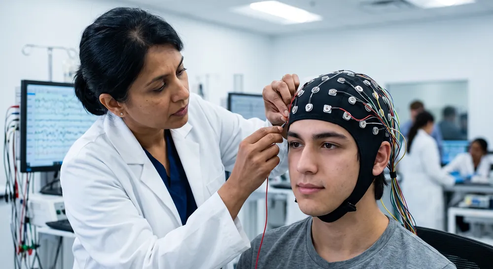
An EEG sensor is an electrode or electrode assembly used to detect electrical signals at the scalp for electroencephalography. These recordings reflect the combined activity of many neurons rather than the activity of a single cell. Sensors are part of a larger system that also includes signal amplification, recording, and analysis.
Understanding the sensor’s role helps distinguish what the hardware measures from what clinicians or researchers may infer from a recording.
How EEG Sensors Work
Neurons communicate through electrical and chemical processes, and coordinated activity across groups of neurons can produce small voltage changes detectable at the scalp. An EEG sensor makes contact with the scalp and forms part of a measurement path; the recording system compares electrical potentials between electrodes and amplifies the resulting signal. The method is noninvasive, but the recorded waveform is affected by the quality of electrode contact as well as by movement, eye activity, and other sources of interference.
An EEG recording is therefore a time-varying electrical measurement that requires context and appropriate interpretation. Reliable contact matters because unstable contact can contribute noise or make recordings harder to compare. Researchers and clinicians also document electrode positions and recording conditions so the data can be interpreted responsibly.
Types of EEG Sensors
EEG sensors are often described by how they contact the scalp and whether a conductive medium is used. Wet electrodes commonly use gel or another conductive medium, while dry designs are intended to record without that preparation step. Semi-dry approaches occupy a middle ground, and sensor materials and shapes vary by system. Our guide to electrode interface explains why electrode selection and placement are closely linked to recording conditions.
The broad distinctions below are useful for an initial comparison, though the performance of a specific system depends on its full design and use:
Sensor approach | Contact method | Practical consideration |
|---|---|---|
Wet | Uses conductive gel or another medium | Requires preparation and cleanup |
Dry | Contacts the scalp without conductive gel | Fit and contact can be important considerations |
Semi-dry | Uses a limited conductive medium | Balances preparation needs with scalp contact |
Integrated cap | Holds electrodes in a planned arrangement | Can support repeatable placement across sessions |
These categories do not by themselves indicate whether a sensor is appropriate for a particular study. Electrode placement, the recording hardware, and the task being studied all affect what can be learned from the data. A consistent placement scheme is especially useful when recordings need to be compared across people or sessions.
How an EEG Sensor Converts Ionic Currents into Electronic Signals
When populations of cortical neurons become synchronously active, they generate extracellular current flows that propagate through the cerebrospinal fluid, skull, and scalp. At the scalp surface, this ionic movement can be detected as a minute potential difference between two locations.
A neuroscience sensor must convert this ionic signal into an electron-based current that recording electronics can handle, a process that occurs through a reversible electrochemical reaction at the electrode–electrolyte interface. The fidelity of that conversion is governed largely by the impedance of the contact: a low and stable impedance ensures that the tiny ionic currents are not attenuated or corrupted by thermal noise before they can be sensed.
When a sensor rests against the skin, it forms a half-cell that develops a characteristic electrical potential. Any mechanically induced disturbance at the interface, such as a patient turning the head or a cable tug, will cause fluctuations in that potential, which can easily overwhelm the microvolt-level brain signals.
For this reason, an ideal EEG sensor must maintain an electrically quiet interface while providing a mechanically stable connection despite the presence of hair and the natural movement of the body. The three broad families of EEG sensor designs—wet, dry, and active—each tackle this transduction challenge with a fundamentally different physical strategy.
Sensor Type | Core Strategy |
|---|---|
Wet | Ionic gel bridge |
Dry | Mechanical hair displacement |
Active | On-sensor buffer amplifier |
Benefits and Limitations of Wet EEG Electrodes
The gold-standard sensor for clinical and research EEG remains the silver/silver chloride (Ag/AgCl) electrode used in conjunction with an electrolytic paste or gel. Its dominance is rooted in the well-characterized electrochemistry of the Ag/AgCl couple.
A silver substrate coated with a layer of silver chloride participates in a reversible redox reaction when placed in contact with a chloride-containing electrolyte: Ag ↔ Ag⁺ + e⁻. This reaction provides a non-polarizable interface, meaning that current can pass across the boundary without a large buildup of overpotential that would distort the recording. The half-cell potential is stable, and the reaction kinetics are fast enough to faithfully track the biologically relevant frequency components of the EEG, typically from below 1 Hz to roughly 50 Hz.
In practice, the metal pellet alone cannot form a reliable electrical path to the scalp. This is where the conductive gel fills a vital role.
Gel is an ionic bridge as it hydrates the outermost dead-cell layer of the skin, the stratum corneum, causing it to swell and reducing its normally high resistance from the megohm range down to a few kilohms. The gel conforms to microscopic pits and furrows in the skin surface, increasing the effective contact area and creating a low-impedance, low-noise pathway for ionic flow.
Because the gel is a viscous fluid, it decouples mechanical motion; the metal pellet can shift slightly inside the gel column without transmitting that displacement as a pressure change at the skin interface. The result is a sensor with contact impedances typically on the order of a few kilohms, low intrinsic noise, and excellent resistance to motion artifact.
These electrical advantages, however, come at a mechanical and practical cost:
Gel dehydration over time raises impedance and eventually causes electrode failure.
Skin preparation requires alcohol cleaning and occasional abrasion, adding time and discomfort.
Gel residue in hair demands post-recording washing, making prolonged unsupervised use impractical.
For any application extending beyond a single day or requiring truly unsupervised wearable consumer EEG headsets, the classic wet electrode becomes impractical. As one recent perspective review notes, while wet electrodes provide the reference standard, the need for “dry scalp electroencephalography sensors as non-invasive probes with high sensitivity, accuracy, comfort and robustness for prolonged and repeated use” has become a primary driver for materials innovation.
How Silicone Dry Electrodes Achieve Gel Free EEG Recording
Dry electrodes abandon the electrolyte gel entirely. Instead of an ionic bridge, the solid conductive sensor surface must be pressed directly against the skin.
The challenge is twofold: electrical and mechanical.
An engineering study attempted to solve this mechanical problem with a novel compliant mechanism that actively displaces hair through a controlled, multiphasic motion. The sensor is fabricated from biocompatible silicone via injection molding, a manufacturing process that enables precise, repeatable production of the flexible structures required.
Unlike a simple linear press, the mechanism executes a screw-like, helical rotational movement. As the actuator advances, the sensor element rotates while simultaneously extending, effectively “corkscrewing” through the hair layer until its tip achieves direct mechanical contact with the scalp. This design aims to provide a stable, reproducible interface without the need for any skin abrasion or fluid couplant.
The study’s functional demonstrators were tested under controlled pneumatic pressure. At an applied pressure of 650 millibars, the prototype achieved a height extension exceeding 3 millimeters and a rotation angle greater than 45 degrees, which met the preset design targets necessary to penetrate the hair layer. The experimental results were compared against a computational model used to design the geometry.
The measured height change deviated from the simulated value by approximately +17%, and the rotational angle deviation was +6%. These discrepancies suggest that while the computational model captures the dominant mechanics, real-world factors such as frictional variations and hair density introduce measurable differences that must be accounted for in any high-precision application.
The silicone compliance is critical here: by distributing pressure over a slightly larger area and allowing the tip to gently flex as it advances, the mechanism prevents the intense point-loading that makes rigid dry electrodes so uncomfortable.
Yet the electrical trade-off remains. Even after the hair has been displaced and mechanical contact has been established, the dry conductive tip—whether a metal coating, a conductive polymer, or a sintered composite—faces the skin without a gel bridge.
The contact impedance is substantially higher than that of a wet electrode, typically measured in the megohm range. At these impedance levels, the sensor becomes extraordinarily sensitive to electromagnetic interference and cable-borne noise, which is where on-sensor electronics become essential.
The Importance of Active Shielding and On-Sensor Electronics
A dry electrode with megohm-level contact impedance connected to a long, unshielded cable forms an excellent antenna. Cable capacitance and the ubiquitous 50 or 60 hertz mains-frequency electric field couple into the high-impedance source, and because of the voltage-divider effect, most of the interfering voltage drops across the electrode interface rather than being attenuated by the amplifier input. Under such conditions, the microvolt-level brain signal can be entirely swamped by interference long before it reaches a traditional central amplifier.
The engineering fix is to move the first stage of amplification as close to the point of transduction as physically possible. Instead of merely placing a passive metal contact against the skin, the sensor element can incorporate a compact, high-input-impedance buffer amplifier directly on the sensor substrate.
One relevant design study details a special electrode chip that integrates an extremely high input impedance stage alongside a superior common mode rejection ratio (CMRR). By buffering the signal at the source, the chip transforms the high-impedance interface into a low-impedance output that can drive a cable with far less susceptibility to electrical noise.
The high input impedance ensures minimal loading of the already-weak ionic currents, while the high CMRR rejects interference that is common to the two differential inputs of a typical EEG channel. Importantly, this active sensor approach does not lower the skin–electrode impedance itself; instead, it electrically decouples that impedance from the rest of the system, so that the signal integrity depends on the local buffer’s performance rather than on the vagaries of cable routing.
This active front-end architecture retains one essential characteristic: it remains a purely electrical transducer. The underlying ionic-to-electronic conversion still occurs at the dry metal–skin interface with whatever half-cell potential and noise that interface produces.
The buffer amplifies and impedance-converts the resulting voltage but cannot suppress low-frequency drifts that arise from the inherently unstable half-cell potential of a dry contact. For this reason, active dry electrodes often require careful material selection for the contact surface—such as gold, platinum, or specialized conductive coatings—that minimize spontaneous potential shifts while maintaining biocompatibility.
Advanced Materials and Multi Modal Frontiers in EEG Sensing
Looking ahead, the sensor substrate itself is expected to evolve beyond standard metals. The same perspective review that identified the push toward dry electrodes also highlights the promise of biocompatible nanomaterials: conductive polymers, graphene flakes, carbon nanotube networks, and hybrid composites that merge high electrical conductivity with elastomeric flexibility.
Such materials offer three potential advantages:
First, their effective surface area can be orders of magnitude larger than a smooth metal pellet, which directly reduces the contact impedance through a purely capacitive mechanism.
Second, their mechanical compliance allows the sensor to conform to the curved topography of the scalp, increasing the true contact area without requiring applied pressure to flatten the skin.
Third, nanomaterial coatings can be engineered to have a lower contact potential drift than bare metals when placed against skin, which may reduce the low-frequency noise that currently limits dry-electrode performance.
The same review proposes a further step by integrating multiple sensing modalities within a single transducer element. Rather than relying solely on an electrical potential measurement, the sensor could incorporate an optical component, such as a miniature photodiode, that simultaneously probes local tissue hemodynamics.
The conceptual reasoning is that because the electrical and optical signals arise from different but related aspects of neural activity, cross-validating them within the same device could increase measurement confidence and help identify artifacts. This approach is described as a form of “redundancy” that probes the same neural information through different physical mechanisms. Noteworthy, it remains, at present, a laboratory concept.
Choosing the Right EEG Sensor
Choosing an EEG sensor begins with the intended use, not with a single specification such as electrode count. A clinical examination, a laboratory study, a classroom demonstration, and a consumer interaction can require different procedures and equipment. Comfort, preparation time, placement repeatability, and compatibility with the acquisition hardware also shape the practical choice.
Therefore, a sensor is only one component of a complete recording system.
EEG Sensor Cost and Factors to Consider
Costs vary with the sensor design and with the rest of the system, including the amplifier, cap or mounting method, software, and accessories. A low purchase price does not necessarily mean a lower total cost if setup materials or replacement components are needed. Conversely, a higher channel count is not automatically more useful; the number and placement of electrodes should fit the question being studied.
A useful comparison considers both capabilities and the conditions in which the system will be used. For example, repeatable placement may be central to a study, while a short educational demonstration may prioritize simple setup. Common factors to compare include:
Electrode type and the preparation it requires.
Number and placement of recording channels.
Compatibility with the amplifier, software, and data format.
Comfort, cleaning, maintenance, and replacement needs.
Taken together, these factors help explain why there is no universally best EEG sensor. Selection should reflect the recording protocol and the level of evidence needed, rather than a feature list in isolation. For medical evaluation, equipment choice and interpretation belong within appropriate clinical practice.
Matching Emotiv Headsets and Sensor Types to Your Application
The contact physics described above explains why Emotiv’s headsets make a deliberate trade-off between signal stability, setup time, channel count, and wearability. The sensor type that suits a 32-channel laboratory study is rarely the one that suits an all-day wellness application, and the right choice depends on the question being asked.
Gel sensors: Emotiv Flex 2 Gel
Flex 2 Gel uses ring-shaped sintered silver/silver chloride (Ag/AgCl) sensors with conductive gel, the same gel-bridge principle that makes wet electrodes the reference standard. The cap offers 74 openings at standard 10-20 positions, and up to 32 sensors can be placed in any of them.
Its strength is stable, low-impedance contact that holds up over long recordings, which makes it the strongest fit for extended sessions, high-density EEG research, and studies where signal stability matters most. The trade-off is the longest preparation time in the lineup, along with the gel residue described earlier.
Even so, setup remains simpler than with a traditional wired laboratory system.
Saline sensors: Emotiv Flex 2 Saline and Epoc X
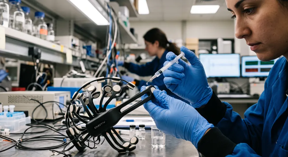
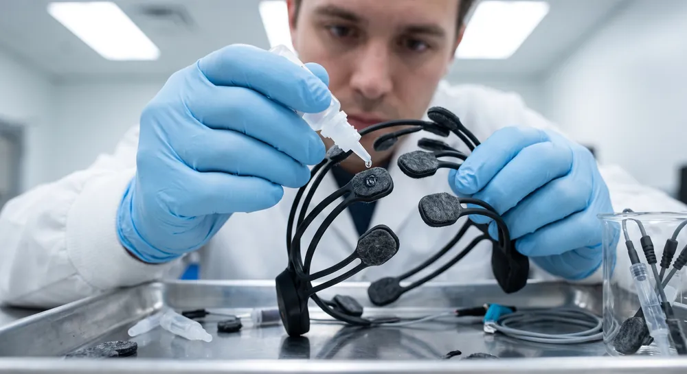
Saline sensors use felt pads soaked in a saline solution to conduct signals between scalp and sensor. They sit between gel and dry designs: no gel to wash out, and setup that takes minutes.
Flex 2 Saline carries the same 32-channel, 74-opening cap, with refill openings that let researchers rehydrate sensors during a session. It suits research that needs a custom montage and a faster workflow than gel allows.
The Emotiv Epoc X is a 14-channel headset with fixed positions at AF3, F7, F3, FC5, T7, P7, O1, O2, P8, T8, FC6, F4, F8, and AF4. Its sensors can be refilled without removing the headset, and its rotating headband and wireless design suit mobile use.
That makes it a strong choice for cognitive research, brain-computer interface (BCI) development, and field studies outside the lab. The trade-off with any saline sensor is that the felts dry over time, which is why refillable designs matter.
Semi-dry sensors: Emotiv Insight
The Emotiv Insight uses hydrophilic semi-dry polymer sensors, which avoid gel and remove most of the preparation burden. It records five channels (AF3, AF4, T7, T8, and Pz) at 128 samples per second.
Its strength is speed. Insight can be on and recorded within minutes, in a lightweight form that suits rapid deployment, introductory BCI projects, developer work, and personal brain-measurement.
Dry sensors: Emotiv MN8
MN8 uses dry conductive elastomer earbud sensors positioned in the left and right ear canals, with two channels plus four references. It is built for discretion and convenience, with minimal setup, up to six hours of battery life, and stereo audio. This suits focus tracking, relaxation, and everyday cognitive wellness, where people want brain data without wearing a cap.
Takeaway
Flex 2 Gel (gel, up to 32 channels): long recordings, high-density research, maximum stability.
Flex 2 Saline (saline, up to 32 channels): configurable research with faster setup.
Epoc X (saline, 14 channels): mobile research, BCI development, field studies.
Insight (semi-dry, 5 channels): rapid deployment, introductory projects, personal use.
MN8 (dry, 2 channels): discreet, everyday cognitive wellness measurement.
No sensor is best across the board. The practical approach is thinking how much signal stability and spatial detail a study needs, and how much setup time it can afford. For raw EEG access and export across any of these headsets, EmotivPRO provides recording, event markers, and quality measurement, and raw data access requires a PRO license.
Why Sensor Contact Physics Matters More Than the Headset Design
The path from brain signal to readable EEG runs through a tiny material boundary where the sensor meets the scalp.
Wet electrodes with conductive gel remain the reference standard because they create low-impedance, electrochemically stable contact, but their need for skin preparation and drying gel restricts long-term wearable use. Dry designs solve the practical problem of hair and gel by using mechanical motion and on-sensor amplifiers, yet their higher contact impedance and unproven real-world durability mean they cannot yet replace wet electrodes.
The most honest takeaway is to separate engineering promise from clinical proof. The silicone corkscrew mechanism, active buffer chips, and nanomaterial coatings are all supported by prototype studies and conceptual reviews, not by large trials across diverse users.
Until future research tests comfort, noise, and multi-day stability head-to-head against wet electrodes, these advances should be viewed as promising pathways rather than validated upgrades.
References
Iacopi, F., & Lin, C. T. (2022). A perspective on electroencephalography sensors for brain-computer interfaces. Progress in Biomedical Engineering, 4(4), 043002.
Griebel, S., Fiedler, P., Streng, A., Haueisen, J., & Zentner, L. (2010). Neuartiger nachgiebiger Mechanismus zur Platzierung trockener EEG-Elektroden über eine Schraubenbewegung. In Biomedical engineering: joint journal of the German Society for Biomedical Engineering in VDE and the Austrian and Swiss Societies for Biomedical Engineering (Vol. 55, No. Suppl. 1, M, pp. 246-249). https\://doi.org/10.1515/BMT.2010.713
Li, G. (2007). Design of a Special Electrode Chip for the Extraction of Electroencephalogram Signal. Journal of Huaqiao University.
Frequently Asked Questions
What is the basic job of an EEG sensor?
An EEG sensor acts as a physical transducer that converts ionic currents from the brain into electronic voltages that recording equipment can read. It must do this faithfully at the point of contact with the scalp, while dealing with obstacles like high skin resistance and hair.
Why is the outer layer of the skin a problem for EEG recording?
The stratum corneum, or outer dead-skin layer, has an intrinsically high electrical resistance, which makes it hard for tiny brain signals to pass through. Additionally, hair physically holds a rigid sensor away from the conductive skin beneath, creating both electrical and mechanical challenges.
How does a traditional wet electrode work?
A wet electrode uses a silver/silver chloride (Ag/AgCl) metal pellet covered by a conductive gel. The gel hydrates and swells the dead outer skin, lowering its resistance, while the Ag/AgCl provides a stable electrochemical reaction that converts ionic current into electronic current.
Why are wet electrodes not ideal for long-term or wearable use?
The gel in wet electrodes dries out over time, which raises impedance and causes the signal to fail. They also require skin preparation and leave residue that demands washing the hair, making them impractical for unsupervised, multi-day use.
What is the main difference between a dry electrode and a wet electrode?
A dry electrode has no gel and must press its solid conductive surface directly against the skin, leading to much higher contact impedance. Without a gel bridge, the dry sensor must also rely on mechanical design—such as compliant silicone structures—to get past hair and maintain a stable interface.
Why is an "active" sensor design important for dry electrodes?
Active sensors place a buffer amplifier directly on the electrode, converting the high-impedance signal into a low-impedance output right at the source. This prevents cable noise and mains interference from swamping the tiny brain signal, even though it does not reduce the actual skin–electrode impedance.
What advantages do nanomaterials offer for future EEG sensors?
Nanomaterials like conductive polymers and graphene can greatly increase the effective surface area, which lowers contact impedance through capacitive coupling. They also flex to conform to the scalp, improving contact without heavy pressure, and may show less potential drift, reducing low-frequency noise.
What is the role of the gel in reducing skin resistance?
The gel acts as an ionic bridge that hydrates the outer stratum corneum, causing it to swell and lowering its normally high electrical resistance. It also fills tiny surface irregularities, increasing contact area and forming a low-impedance, low-noise pathway for ionic current.
Emotiv is a neurotechnology leader helping advance neuroscience research through accessible EEG and brain data tools.
Medical Disclaimer: The information provided on this website is for educational and informational purposes only and is not intended as medical or health advice. This content may contain errors and should not be relied upon to make life-altering health, medical, or lifestyle choices. Always seek the advice of your physician or other qualified health provider with any questions you may have regarding a medical condition or treatment.
Christian Burgos