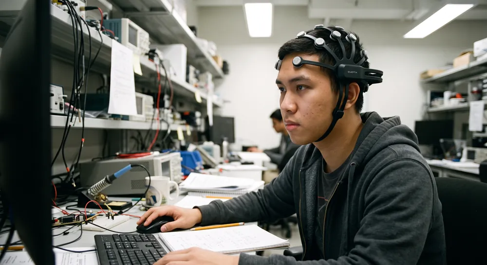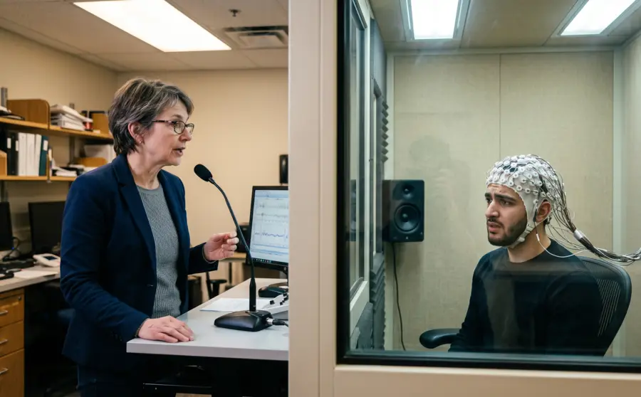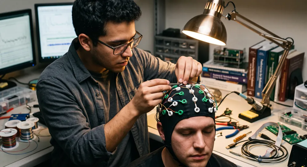
Measuring the brain’s electrical activity from outside the skull offers a direct, real-time view of neural processing without surgical intervention. An electroencephalogram, or EEG, captures these faint voltage fluctuations through sensors placed on the scalp.
Yet for decades, the practical act of positioning those sensors remained one of the field’s most stubborn bottlenecks. In traditional high‑density setups, a technician must locate dozens of landmarks on a participant’s head, scrub each small area, apply conductive gel, and secure each electrode individually. The process can take 30 minutes or more, requires repeated precision, and introduces spatial inconsistencies between sessions that complicate data comparisons.
The EEG cap offers a fundamentally different approach. Rather than treating each electrode as a separate component, the cap integrates all sensors into a single, pre‑assembled fabric or polymer structure that can be pulled onto the head like a fitted hat. This transforms the setup from a tedious, error‑prone procedure into a rapid, repeatable process.
The following sections examine the structural design, functional capabilities, and research‑backed evidence for integrated EEG caps.
What is an EEG Cap?
An EEG cap is a fabric or flexible head covering fitted with electrode holders at standardized scalp locations. During electroencephalography, the electrodes detect very small voltage differences produced by synchronized neural activity, while recording equipment amplifies and stores those signals.
Cap-based placement makes setup more repeatable than positioning every electrode independently. It does not, by itself, interpret the recorded activity or establish a diagnosis.
How Does an Electrode Cap for EEG Work?
An electrode cap for EEG follows a placement map, often based on the international 10-20 system. Measurements between anatomical landmarks guide the location of electrodes so that recordings can be compared across sessions and, with appropriate care, across participants. The standardized 10-20 system provides a common naming and placement framework, although denser systems may be chosen when more spatial information is needed.
Before recording, the electrodes must make suitable contact with the scalp. Depending on the design, this may involve conductive gel, saline, conductive fabric, or spring-loaded contacts. The amplifier measures voltage differences between channels and a reference, while software displays the resulting waveforms for clinical or research interpretation.
Good placement is only one part of a reliable recording. Cable movement, muscle activity, eye movements, sweat, and poor contact can introduce artifacts that resemble or obscure neural signals. Thus, careful preparation, impedance checks where applicable, and accurate documentation of channel locations support more trustworthy EEG data.
Types of EEG Caps
EEG caps vary mainly in electrode technology, preparation method, materials, and intended setting. Some prioritize stable low-impedance contact, while others reduce setup time and avoid conductive paste. The best design depends on the recording protocol, participant population, duration, and degree of movement expected during the session.
Wet EEG Caps
Wet EEG caps use a conductive medium, commonly gel or saline, between the electrode and scalp. The medium helps reduce impedance and can provide a stable connection when the cap is fitted and prepared correctly. These systems are widely used when signal quality and established recording procedures take priority over rapid cleanup.
Preparation can take longer because hair may need to be parted and each contact checked. Gel can also feel cool or require washing after the recording. Even so, wet systems remain useful for recordings in which stable contact over many channels is more important than convenience.
Dry EEG Caps
Dry EEG caps use electrodes designed to contact the scalp without applying conductive gel. Their principal practical advantage is reduced preparation and cleanup, which can be valuable in repeated sessions or settings where participants need to be fitted quickly. Some dry electrodes use flexible or spring-loaded structures to accommodate differences in hair and scalp shape.
Signal quality depends strongly on electrode pressure, hair thickness, movement, and the specific contact design. A dry system is not automatically easier for every participant; firm contacts may be noticeable, and some hair types can make scalp contact difficult. Testing the cap with the intended population is therefore part of evaluating its suitability.
EEG Skull Cap and Hybrid Cap Designs
The terms EEG skull cap and hybrid cap often describe head-worn designs that combine a close-fitting textile structure with different electrode or sensor arrangements. A hybrid design may use wet contacts in some positions and dry or semi-dry contacts in others, depending on the recording objective. These arrangements seek a practical balance between contact quality, setup time, and comfort.
The cap must remain stable without creating excessive pressure. Chinstraps, adjustable bands, flexible fabrics, and carefully routed cables can reduce movement, but each added component affects fitting and cleaning. For longer sessions, a modest reduction in preparation time may matter less than maintaining comfort and consistent electrode contact.
EEG Cap Design: Inflatable vs. Adjustable Structures
The core engineering challenge of an EEG cap is to keep multiple dry sensors in stable, low‑resistance contact with the scalp even through hair, across diverse head morphologies, and without the messy conductive pastes that wet electrodes require. Two prototype systems illustrate distinct solutions.
One cap, the inflatable wireless system described by Yu et al., integrated 32 spring‑loaded dry sensors into a ventilated fabric shell. Beneath the outer layer, a novel gasbag structure can be inflated to push the sensor tips gently but firmly against the scalp.
This inflation mechanism adapts to the curvature of the head and helps displace hair from the electrode‑skin interface. Because the gasbag exerts distributed pressure rather than relying on the elasticity of the cap alone, contact quality becomes less dependent on head size.
The cap also houses a miniature circuit board, a rechargeable battery, and a Bluetooth transmitter, eliminating the need for a trailing cable bundle. All electronics and the power source reside within the cap itself, making the unit fully wearable and self‑contained.
On the other hand, a 2019 study by Wu et al. presents a cap that takes a different path to fit and electrode alignment. Its “concise structure” uses a lightweight frame with independent size‑adjustment mechanisms in both horizontal and vertical axes. This means the cap can be expanded or contracted to suit different head circumferences and crown heights.
Crucially, the dry electrodes are mounted in the cap but are not rigidly fixed in the vertical plane; each one can be adjusted independently to press down until it achieves adequate contact. So a subject with a highly uneven scalp surface does not force the entire cap to tilt—each sensor can find its own mechanical equilibrium. The electrodes integrate into the cap’s overall geometry, yet they retain the ability to move relative to the shell.
Both designs converge on a single, critical property: the electrodes remain locked in a predetermined spatial arrangement. Unlike manual placement, where a technician might inadvertently shift one electrode a centimeter off its intended landmark, the cap’s pre‑designed layout fixes inter‑electrode distances.
This invariant relative geometry is what gives the integrated cap its power for spatial mapping of brain signals, because the recorded voltage distribution across the scalp can be referred to a consistent set of coordinates every time the cap is worn.
Why EEG Caps Enable Faster and More Consistent Electrode Placement
The practical advantage of the integrated cap becomes clear when setup time is measured against the traditional single‑electrode method. The adjustable cap study specifically reports that the device can be “rapidly worn and adjusted,” emphasizing that the entire array can be donned as a single unit and then fine‑tuned in seconds.
In a manual workflow, a technician places each electrode, checks impedance, repositions it, and repeats that cycle for every channel. That sequence multiplies the opportunity for spatial error. With the cap, the gross positioning is accomplished in one motion, and only small individual adjustments are needed.
In addition, the inflatable cap study underscores that the fully integrated wireless system—sensors, amplifier, battery, transmitter—makes the setup practical for experimental use without needing to tether the participant to a recording station. The subject can simply put on the cap, the gasbag is inflated to optimize contact, and recording begins.
This not only speeds up the preparation but also reduces the physical discomfort and social awkwardness of having a technician’s hands on the head for a prolonged period, which can be especially helpful when working with children, clinical populations, or individuals with sensory sensitivities.
Beyond speed, the fixed electrode layout addresses a more subtle but significant limitation of manual placement: session‑to‑session variability. Even well‑trained operators will introduce millimeter‑scale differences in electrode position when reapplying sensors on different days.
These small shifts can change the amplitude and topography of the recorded waveforms enough to blur subtle experimental effects or make longitudinal monitoring unreliable. By mechanically enforcing the same inter‑electrode spacing every time, an integrated cap reduces this source of variance.
The result is a recording setup that yields more comparable data across subjects and across repeated measurements from the same individual, enabling more confident conclusions about genuine neurophysiological changes rather than artifacts of placement.
From Prototype to Practice: The Emotiv Flex 2
The Emotiv Flex 2 is a 32-channel wireless EEG head cap built around configurable sensor placement rather than one fixed electrode array.
Rather than assigning each of its 32 sensors to a permanently fixed scalp location, the Flex cap provides 72 mounting positions laid out according to the standard 10-20 system, so the same hardware can be reconfigured into different channel montages depending on the study's needs. Caps are also offered in multiple head-circumference sizes (54, 56, and 58 cm as standard, with additional sizes available on request), addressing the fit variability discussed throughout this piece.
The system is available with either gel or saline-based sensors, placing it across both the wet and semi-dry categories described earlier. The saline sensors use felt pads that can be re-wetted and sterilized between uses rather than requiring a fresh gel application each session, which shortens cleanup time relative to a traditional gel-based setup.
Wireless transmission and real-time, per-channel contact-quality monitoring are built into the system, giving a continuous readout of electrode contact during a recording rather than relying only on a pre-session impedance check–a version of the automated contact monitoring this article points to later as a likely direction for future cap designs.
This kind of system has also been evaluated directly against a laboratory benchmark. For instance a peer-reviewed study compared a saline-sensor Flex cap to a wired, research-grade EEG system across five established paradigms, including mismatch negativity, P300, and resting-state recordings. The two systems produced statistically equivalent MMN and P300 waveform components, with high correlation between the recorded waveforms, and similar patterns of resting-state alpha power.
Applications of EEG Caps
EEG caps support several forms of measurement, from routine clinical recordings to controlled laboratory experiments. The cap standardizes electrode placement, but the meaning of the resulting waveform depends on the protocol, participant state, recording quality, and interpretation method. EEG is one tool within broader neuroscience, rather than a complete account of brain function on its own.
Medical Diagnostics
In clinical settings, EEG recordings can help assess patterns of electrical activity associated with neurological conditions. They may be used alongside medical history, examination, imaging, and other tests when evaluating events such as suspected seizures or altered awareness. The cap provides a repeatable arrangement of electrodes, while trained professionals interpret the recording in context.
Notably, a cap cannot determine a diagnosis independently. Normal or abnormal findings may have different significance depending on timing, sleep or wakefulness, medications, artifacts, and the clinical question. For that reason, the recording procedure and its interpretation are treated as connected but distinct parts of an evaluation.
Neurofeedback and Brain Training
In neurofeedback research and practice, EEG signals may be displayed or translated into feedback linked to a participant’s ongoing activity. A cap can make repeated electrode placement more consistent across sessions, which helps reduce variation caused by changing sensor locations. The usefulness of feedback still depends on signal quality, protocol design, and an appropriate interpretation of the measured signal.
Common practical concerns include movement, eye activity, muscle tension, and changes in contact during a session. These sources can influence the measured waveform and may be mistaken for changes in the target activity. Training protocols therefore require careful monitoring rather than assuming that every visible fluctuation reflects a meaningful neural change.
Research and Development
Researchers use EEG caps to study perception, attention, sleep, sensory processing, motor activity, and responses to controlled stimuli. Higher channel counts can provide denser spatial sampling, while lower-density arrangements may be more appropriate for portable or repeated measurements. Event timing, sampling rate, referencing, and artifact handling are as important as the cap itself.
When comparing results, researchers document electrode locations, cap size, reference choices, contact conditions, and missing channels. These details make data easier to reproduce and interpret.
A carefully selected cap can support the experiment, but it cannot compensate for an unclear research question or incomplete recording metadata.
Choosing the Right EEG Cap
Choosing an EEG cap begins with the recording objective rather than the channel count alone. Clinical monitoring, laboratory experiments, mobile studies, and repeated training sessions impose different demands on preparation, stability, portability, and cleaning. The intended participants also matter, since hair, head shape, age, and tolerance can affect contact quality.
A useful comparison considers the full workflow: fitting, skin preparation, impedance checking, cable management, removal, and sanitation. A cap that is quick to place may require more attention to pressure or hair interference, while a system with more preparation may offer a familiar and stable recording process. Signal quality depends on the whole setup, not simply on the electrode material.
The following factors help organize a technical comparison without treating any one feature as decisive:
Electrode technology and whether conductive medium is required.
Number and geometry of channels for the planned protocol.
Fit range, adjustability, and pressure distribution.
Cleaning, replacement, and maintenance requirements.
Compatibility with the amplifier, reference arrangement, and recording software.
EEG Head Cap Materials and Electrode Density
Materials influence how an EEG head cap feels, stretches, cleans, and maintains its shape. Common cap structures may use textiles, elastic fibers, mesh, silicone-like components, or molded supports, while electrodes may be made from conductive metals, coated contacts, or conductive composites. No material is ideal for every scalp, hair type, duration, or cleaning process.
Electrode density is a separate design decision. A low-density arrangement can simplify preparation and reduce participant burden, whereas denser layouts sample more scalp locations and may support finer spatial analyses. The added electrodes also create more opportunities for poor contact, cable tangling, and data-quality checks.
A practical comparison can be framed around the relationship between construction and measurement needs:
Design factor | Lower-density or simpler design | Higher-density or more structured design |
|---|---|---|
Preparation | Usually faster and less complex | Often requires more placement and contact checks |
Spatial sampling | Broad coverage with fewer locations | More detailed sampling across the scalp |
Comfort | Fewer contact points and lighter routing may help | More contacts and hardware may increase pressure |
Data handling | Smaller channel set and simpler review | Larger data volume and more artifact management |
The table describes tendencies rather than fixed rules. A well-fitting dense cap may be more comfortable than a poorly fitting simple one, and material durability can matter as much as the initial design for facilities conducting frequent recordings.
The Future Role of Integrated EEG Caps in Neuroscience
Future EEG caps are likely to focus on making recordings easier to repeat outside highly controlled laboratories. Wireless transmission, lighter materials, flexible electronics, and improved contact monitoring may reduce the burden of cables and preparation. Such developments must still preserve accurate timing and stable signal acquisition.
Higher-density systems may also become more manageable through miniaturized electronics and improved software. More electrodes can provide richer spatial sampling, but they also increase data volume, setup demands, and the need for careful artifact management. Hardware progress therefore needs to be matched by clear analysis methods and complete metadata.
Another direction is more adaptive fitting for different head shapes, hair types, and age groups. Automated checks could identify poor contact or shifting electrodes during a session, while modular designs could allow researchers to change sensor layouts without replacing the entire cap. These advances may broaden use, but validation remains necessary before a new design is treated as interchangeable with established systems.
Why Integrated EEG Caps Are a Promising Step for Practical Neuroscience
Integrated EEG caps turn electrode setup from a slow, error-prone ritual into a single wearable step, making high-density brain recording more practical for research and brain-computer interface work. By holding electrodes in a fixed spatial arrangement, they also reduce session-to-session variability and support more consistent comparisons across people and over time.
Future work with larger, more diverse populations and direct benchmark comparisons will determine where caps truly fit. For now, the integrated EEG cap is a genuine but modest engineering advance whose full role in research and clinical practice is still being defined.
References
Yu, Y. H., Lu, S. W., Chuang, C. H., King, J. T., Chang, C. L., Chen, S. A., ... & Lin, C. T. (2016). An inflatable and wearable wireless system for making 32-channel electroencephalogram measurements. IEEE Transactions on Neural Systems and Rehabilitation Engineering, 24(7), 806-813. https://doi.org/10.1109/TNSRE.2016.2516029
Wu, X., Zheng, L., Jiang, L., Huang, X., Liu, Y., Xing, L., ... & Chen, H. (2019). A dry electrode cap and its application in a steady-state visual evoked potential-based brain–computer interface. Electronics, 8(10), 1080. https://doi.org/10.3390/electronics8101080
Williams, N. S., McArthur, G. M., de Wit, B., Ibrahim, G., & Badcock, N. A. (2020). A validation of Emotiv EPOC Flex saline for EEG and ERP research. PeerJ, 8, e9713. https://doi.org/10.7717/peerj.9713/table-6
Frequently Asked Questions
What is an EEG cap and how does it work?
An EEG cap is a pre-assembled fabric or polymer structure that integrates all electrodes into a single unit that can be pulled on like a hat. It replaces the slow process of gluing, gelling, and securing individual electrodes onto the scalp. The cap's internal geometry keeps the electrodes in a fixed and consistent spatial layout.
Why are EEG caps faster to set up than traditional electrode placement?
Traditional setups require a technician to position, scrub, and gel each electrode individually, which is a slow and error-prone process. An EEG cap allows the entire electrode array to be donned in one motion, requiring only small fine-tuning adjustments. This compresses the preparation time into a single don-and-record step.
Why is the fixed electrode layout of a cap important for data quality?
In manual placement, a technician might inadvertently shift an electrode by a centimeter or more off its intended landmark. A cap mechanically enforces the same inter-electrode spacing every time it is worn, eliminating this session-to-session variability. This consistency allows researchers to compare brain signal topographies across different days and across participants with greater confidence.
Why is a cap form factor important for brain-computer interfaces (BCIs)?
A cap consolidates a high-density array of electrodes into a single wearable system, making multi-channel neural recording practical for real-time decoding. In a BCI test, the cap's spatially consistent signals provided enough data for a classification algorithm to accurately identify which target a user was attending to. This bridges the gap between the rich information of multi-electrode recordings and the practical need for a setup that does not collapse under its own complexity.
Emotiv is a neurotechnology leader helping advance neuroscience research through accessible EEG and brain data tools.
Medical Disclaimer: The information provided on this website is for educational and informational purposes only and is not intended as medical or health advice. This content may contain errors and should not be relied upon to make life-altering health, medical, or lifestyle choices. Always seek the advice of your physician or other qualified health provider with any questions you may have regarding a medical condition or treatment.
Christian Burgos