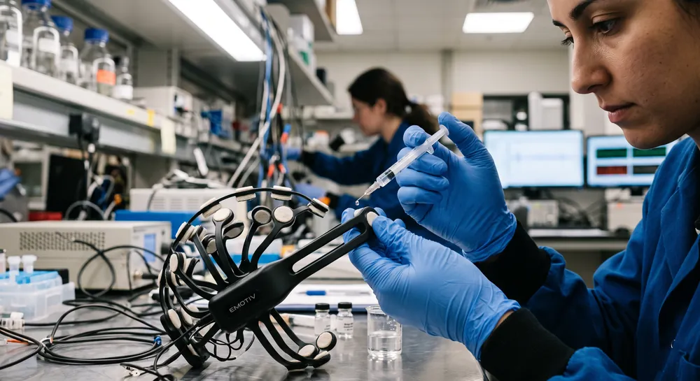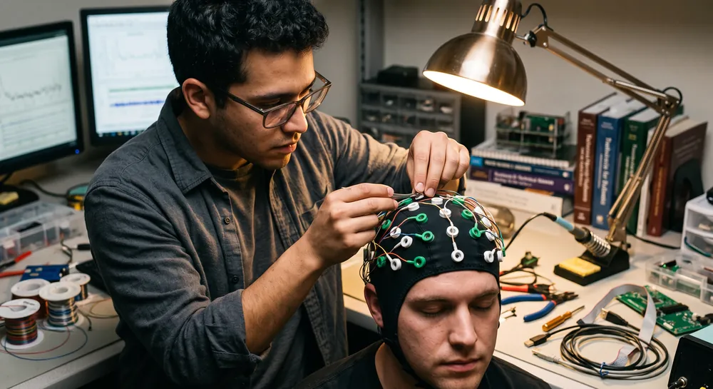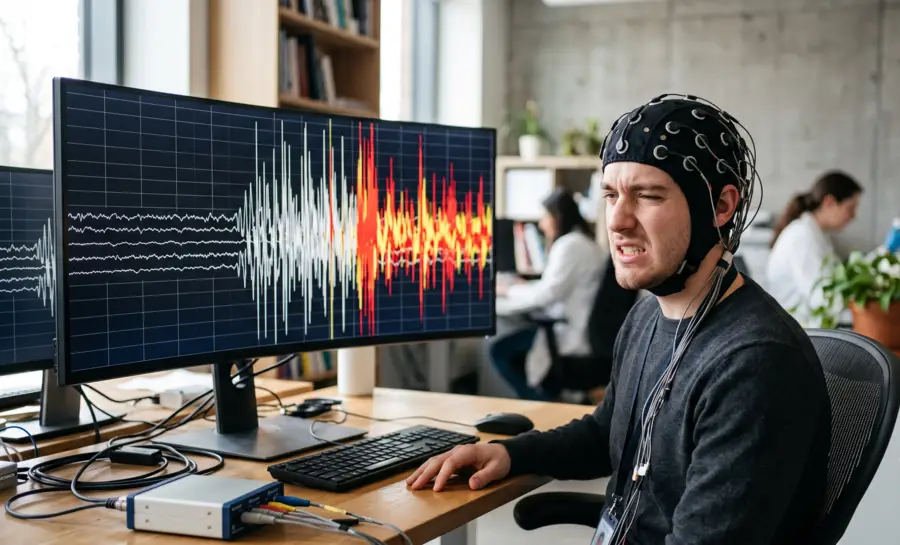
Artifacts are unwanted signals not generated by the brain that can distort the visual interpretation of an electroencephalogram and corrupt the algorithmic analyses that drive brain‑computer interfaces or mental state monitoring.
Whether you are reading a raw EEG trace for epilepsy markers or feeding data into a machine‑learning pipeline, undetected artifacts can masquerade as pathological waveforms or introduce variance that degrades model performance.
This practical field guide walks you through the two broad categories of EEG artifacts, explains how to recognize their distinctive time‑domain signatures, and lays out the manual cleaning steps that remain essential before any computational processing.
The Two Fundamental Categories of EEG Artifacts
Every unwanted fluctuation in an EEG recording arises from one of two origins. Subject‑related, or physiological, artifacts are generated by the participant’s own body. These include eye blinks, muscle tension, heartbeat, and sweat gland activity, all of which produce electrical fields that the scalp electrodes pick up alongside genuine brain activity.
Technical, or non‑physiological, artifacts stem from the recording equipment and environment. Loose electrodes, cable sway, amplifier saturation, and mains electrical interference fall into this second group.
Distinguishing between the two categories is the first skill to master because the prevention and removal strategies differ fundamentally. A muscle burst from a clenched jaw requires a different intervention than a periodic 60‑hertz hum entering through a poorly shielded cable.
How to Recognize Subject‑Related Artifacts
Eye Blinks
The eye behaves like a small electrical battery. The cornea carries a positive charge relative to the retina, forming a corneo‑retinal dipole. When a participant blinks, the eyelid sweeps across this dipole and drags the positive field upward, generating a large, positive voltage deflection at the frontal electrodes nearest the eyes.
In the time‑domain trace, a blink appears as a high‑amplitude, slow wave that reaches its maximum at frontopolar channels such as Fp1 and Fp2, then attenuates sharply as you look toward central or posterior derivations. Because the waveform is so stereotyped, a trained eye can spot it almost instantly.
Eye Movements and Saccadic Spikes
When the eyes move laterally without a blink, the corneo‑retinal dipole rotates rather than being covered. This produces a step‑like shift in the electro‑oculogram signal that may bleed into adjacent EEG channels.
In addition, the rapid contraction of the extraocular muscles during a saccade creates a brief, sharp electrical transient known as the saccadic spike potential. On a raw trace, these spikes can look deceptively similar to epileptiform activity. They are typically confined to frontal and temporal electrodes and coincide with a change in gaze.
Recognizing them prevents a false clinical impression and avoids the unnecessary rejection of otherwise clean neural data. The spike’s brevity, its correlation with a horizontal or vertical eye movement, and its lack of an after‑going slow wave help differentiate it from a genuine cortical discharge.
Muscle Activity
Scalp, face, and neck muscles are prolific generators of high‑frequency electrical noise. When a participant clenches their jaw, furrows their brow, or even holds tension in the neck, motor unit action potentials fire asynchronously and summate into a spiky, irregular interference pattern.
In the time domain, muscle artifact looks like a rapid burst of sharp, low‑amplitude deflections riding on top of the EEG, often most prominent over temporal or frontal electrodes. Its frequency content sits mainly above 20 Hz, well into the beta and gamma bands where many cognitive measures reside, making it particularly troublesome for mental state monitoring and brain‑computer interface applications.
High‑density EEG setups with closely spaced electrodes can help localize the muscle source, but the first line of defense is instructing the participant to relax and providing physical support for the head.
Sweat and Electrodermal Shifts
Changes in skin conductance due to sweat gland activity alter the electrode‑skin interface. This introduces a very slow, smooth drift of the baseline voltage, often below 1 Hz, that can wander across several seconds.
The artifact does not appear as a spike or rhythmic buzz but as a gentle, wave‑like rolling of the entire trace. It is most common during long recordings, in warm environments, or with anxious participants. This low‑frequency confound can be managed by maintaining a stable room temperature and ensuring good electrode contact.
Cardiac Signals
The electrical field of the heart is powerful enough to reach the scalp, especially when electrode impedances are high or when a reference electrode sits near a blood vessel over the mastoid or ear lobe.
The resulting electrocardiogram artifact shows up as a regular, sharp QRS‑complex repeating at the heart rate—roughly once per second. It often bleeds into many channels simultaneously with a consistent timing offset, and classifiers trained on broad‑spectrum artifact features can detect it alongside other contaminants.
If the cardiac waveform is visible but not overwhelming, it can often be subtracted by recording a dedicated ECG channel; otherwise, the affected segments may need manual rejection.
Artifact | Signature |
|---|---|
Eye blink | Frontal slow wave |
Eye movement | Step-like shift |
Muscle | High-frequency spikes |
Sweat | Slow baseline drift |
Cardiac | Sharp QRS complex |
Recognizing Technical Artifacts in the Trace
Electrode Pops and Movement‑Related Spikes
A sudden change in impedance caused by a dried‑out gel bridge, a tugged cable, or a shifting electrode generates an abrupt, steep transient often called a pop. This falls under the “generic discontinuities” category that automated algorithms like ADJUST are designed to flag.
In the time domain, an electrode pop typically looks like an isolated spike of extreme amplitude that may saturate the amplifier, flat‑lining the signal for a moment before a slow recovery. Unlike a biological spike, it is not surrounded by ongoing physiological activity and usually appears on a single channel, making it easy to identify during visual inspection.
A neonatal EEG study by Hagmann et al. similarly documented frequent movement interference that altered the amplitude‑integrated trace, emphasizing that even brief physical disturbances create measurable contamination.
Mains Interference
Capacitive coupling of the electrical supply into the recording setup produces a continuous, rhythmic “buzz” at the mains frequency—50 Hz in Europe and much of the world, 60 Hz in North America.
On a raw EEG trace, this interference appears as a thick sine‑wave superimposed on every channel, often with a consistent amplitude. Even a small amount of line noise can mask low‑voltage cerebral rhythms, and its presence signals a problem with electrode impedance, shielding, or grounding.
It is invariably a technical artifact, not a neural oscillation, and its fixed frequency makes it straightforward to identify by eye or with a simple spectral plot.
Loose Electrodes and Cable Movement
When an electrode is not securely attached to the scalp or its cable sways mid‑recording, the mechanical disturbance introduces irregular low‑frequency fluctuations. These can look like a slow, erratic drift or a burst of high‑amplitude irregular waves on a single channel.
The sporadic nature and confinement to one electrode immediately distinguish this artifact from widespread physiological events such as a blink or a muscle tensing. Annotating such moments in real time saves considerable effort later by narrowing the search for bad data segments.
Preventing and Documenting Artifacts During a Recording
The most effective artifact management strategy begins long before the first data point is saved.
A relaxed participant yields dramatically fewer muscle and eye‑movement artifacts. Therefore, take time to explain the session, provide a comfortable chair with head support, and give the eyes a stable fixation point to reduce spontaneous blinks and saccades. These steps are so foundational that they are rarely spelled out in research papers, yet every study that reports clean data implicitly relies on them.
Electrode application is the next line of defense. Meticulous skin preparation, adequate conductive gel, and careful cable routing minimize both environmental pickup and movement‑related pops.
The aforementioned infant EEG study found that electrical and movement interferences occurred with similar frequency, confirming that no recording environment is inherently immune to technical problems. Checking impedances before and after the session and securing cables to the clothes or chair removes a large fraction of preventable artifacts.
Finally, keep a running log of visible artifacts during the recording. Note the time of any gross movement, cough, or electrode adjustment. This kind of manual annotation mirrors the expert labeling used to train automatic classifiers, where human raters mark segments to serve as ground truth for machine‑learning algorithms.
A simple handwritten timestamp or an event marker in the acquisition software transforms guesswork into reproducible documentation.
Manual Rejection and Interpolation: Cleaning Before Computation
Visual Inspection of the Continuous Trace
Before delegating artifact removal to any signal processing algorithm, scroll through the raw data and use the waveform signatures described above to identify contaminated intervals.
It’s often recommended to look for the characteristic frontal slow wave of a blink, the high‑frequency fizz of muscle activity, the isolated spike of a pop, or the persistent sine‑wave of mains noise. This step trains your eye to distinguish artifacts from genuine brain activity and prevents the automatic rejection of rare, high‑amplitude neural events that might superficially resemble artifacts.
Frontal slow wave: indicates an eye blink
High‑frequency fizz: indicates muscle activity
Isolated spike of extreme amplitude: indicates an electrode pop
Persistent sine‑wave: indicates mains interference
Marking and Excluding Bad Time Segments
Once you have identified a stretch of artifact‑laden data, mark it for exclusion. This might involve logging the start and end times or using the marking tools built into the EEG analysis software.
Manual removal of the worst‑contaminated segments is the standard that even state‑of‑the‑art automated methods built upon. In the work that produced the ADJUST algorithm, for example, experts manually labeled 640 components to create a training set, and the classifier’s performance was then measured against this manually defined ground truth.
Removing the most egregious artifacts by hand reduces the burden on downstream algorithms and prevents them from spreading noise into neighboring channels during spatial filtering steps.
Interpolating a Consistently Noisy Channel
When a single electrode is unreliable for the entire recording because of a loose contact, dried gel, or mechanical instability, removing that channel and reconstructing its data from neighboring clean electrodes preserves the overall data dimensionality.
Interpolation uses the spatial voltage distribution across the scalp to estimate what the bad channel would have recorded, effectively replacing the artifact with a plausible, neutral signal. This step keeps the dataset intact for topographical analyses and avoids the data loss that would come from discarding the channel entirely.
Why Manual Cleaning Remains Essential
Even the most sophisticated automatic classifiers benefit from a dataset that has been pre‑cleaned of gross artifacts. The neonatal study’s finding that artifacts occupied 12% of recording time and influenced clinical interpretation illustrates how easily unremoved contamination can distort conclusions.
Therefore, manual inspection ensures that the largest and most obvious artifacts are gone before the data enter an effective computational pipeline, where they might otherwise dominate the variance and mislead the decomposition. It also builds an intimate familiarity with the dataset that no algorithm can replicate.
How Automated Removal Supports Manual Work
Independent Component Analysis as a Dissection Tool
Once the worst segments are manually removed, ICA can decompose the remaining EEG into statistically independent source components. This technique un‑mixes the mixture of brain and artifact signals picked up by the scalp electrodes, separating eye movements, blinks, and muscle activity into discrete components based on their unique spatial and temporal patterns.
A component with a frontal projection and a waveform that closely matches the blink signal can then be identified and subtracted, leaving the rest of the data neurophysiologically intact. ICA’s strength lies in its ability to handle multiple artifact sources that occur simultaneously, a task that simpler regression‑based correction frequently over‑ or under‑corrects.
The Reliability of Modern Classifiers
Automated methods have now reached a level of performance that closely matches expert human judgment.
One subject‑independent linear classifier, trained on six features drawn from the frequency, spatial, and temporal domains, achieved a mean squared error below 10% on a reaction‑time dataset, which is within the range of disagreement between two human experts.
Furthermore, the ADJUST algorithm, which combines spatial and temporal artifact‑specific features, agreed with manual expert labeling on 95.2% of the data variance when tested on a completely independent dataset, and its removal of artifacted components led to a clean reconstruction of event‑related potentials.
A different approach using autoregressive models combined with a support vector machine classified several types of subject‑generated artifacts across individuals with approximately 94% accuracy.
For eye artifacts specifically, an algorithm that incorporated eye‑tracker information to objectively identify the relevant components performed nearly twice as effectively as human experts who had to rely on component topographies alone, achieving a receiver operating characteristic area under the curve greater than 0.99.
These figures do not replace manual cleaning but demonstrate that the field now has reliable computational assistants that can handle the repetitive, high‑volume identification once done entirely by hand.
Balancing Artifact Removal and Signal Preservation
Aggressive artifact rejection can inadvertently strip away genuine neural information. In a motor imagery brain‑computer interface paradigm, researchers found that they could safely remove up to 60% of the most artifactual ICA components while still preserving the discriminant information the BCI system used to decode the user’s intent.
This finding underscores the crucial principle that not every component that carries some artifact variance is devoid of neural signal. Manual review of the components selected by an automatic classifier remains advisable to confirm that the rejection threshold does not cross into the territory of useful brain activity.
Why Artifact Awareness Determines Data Quality
Artifacts directly influence both visual interpretation and quantitative measurements. Whether the goal is a clinical diagnosis, a neuroscience discovery, or a consumer wellness application, the pathway to reliable results runs through a clear understanding of where artifacts come from, how they look on the raw trace, and the disciplined process of preventing, documenting, and manually removing them.
ICA‑based automation can accelerate the cleaning workflow, but it operates most effectively on data that has already been inspected by a trained human eye. A 12% contamination rate may not sound high, but when that fraction can alter a medical decision or obscure a subtle cognitive biomarker, the investment in artifact management becomes inseparable from the scientific integrity of the recording.
References
Plöchl, M., Ossandón, J. P., & König, P. (2012). Combining EEG and eye tracking: identification, characterization, and correction of eye movement artifacts in electroencephalographic data. Frontiers in human neuroscience, 6, 278. https://doi.org/10.3389/fnhum.2012.00278
Mognon, A., Jovicich, J., Bruzzone, L., & Buiatti, M. (2011). ADJUST: An automatic EEG artifact detector based on the joint use of spatial and temporal features. Psychophysiology, 48(2), 229-240. https://doi.org/10.1111/j.1469-8986.2010.01061.x
Hagmann, C. F., Robertson, N. J., & Azzopardi, D. (2006). Artifacts on electroencephalograms may influence the amplitude-integrated EEG classification: a qualitative analysis in neonatal encephalopathy. Pediatrics, 118(6), 2552–2554. https://doi.org/10.1542/peds.2006-2519
Winkler, I., Haufe, S., & Tangermann, M. (2011). Automatic classification of artifactual ICA-components for artifact removal in EEG signals. Behavioral and brain functions, 7(1), 30. https://doi.org/10.1186/1744-9081-7-30
Lawhern, V., Hairston, W. D., McDowell, K., Westerfield, M., & Robbins, K. (2012). Detection and classification of subject-generated artifacts in EEG signals using autoregressive models. Journal of neuroscience methods, 208(2), 181-189. https://doi.org/10.1016/j.jneumeth.2012.05.017
Frequently Asked Questions
What are the two main categories of EEG artifacts, and how do they differ?
EEG artifacts fall into two categories: subject-related (physiological) artifacts generated by the participant's body—such as eye blinks, muscle tension, heartbeat, and sweat—and technical (non-physiological) artifacts stemming from equipment or environment, like loose electrodes, cable sway, and mains interference. The distinction matters because each category requires different prevention and removal strategies; for example, a muscle burst needs a different intervention than a 60-Hz line hum.
How can you recognize an eye blink artifact on an EEG trace?
An eye blink appears as a large, positive, slow wave that is strongest over frontal electrodes like Fp1 and Fp2, and it quickly fades toward central or posterior channels. Because the cornea carries a positive charge relative to the retina, the eyelid sweeping across this dipole creates a stereotyped, high-amplitude deflection that is easy to spot with training.
Why can muscle activity be especially problematic for brain-computer interfaces?
Muscle artifacts from jaw clenching, brow furrowing, or neck tension produce high-frequency, spiky interference that often overlaps with beta and gamma brainwave bands where cognitive measures and mental state monitoring operate. This contamination can corrupt algorithmic analyses and degrade the performance of brain-computer interface models, making it one of the most troublesome physiological artifacts to manage.
What does a mains interference artifact look like, and what causes it?
Mains interference appears as a continuous, rhythmic sine-wave "buzz" superimposed on every EEG channel, usually at 50 or 60 Hz depending on the region's electrical supply. It arises from capacitive coupling of the power source into the recording setup due to poor electrode impedance, inadequate shielding, or grounding problems, and it is clearly a technical artifact rather than a neural signal.
Why is manual annotation of artifacts during a recording important?
Keeping a running log of gross movements, coughs, or electrode adjustments during the session helps narrow down bad data segments later and transforms guesswork into reproducible documentation. This manual annotation also mirrors the expert labeling used to train automated classifiers, providing a ground truth that improves the reliability of both manual and computational cleaning.
What is the purpose of interpolating a consistently noisy EEG channel?
When a single electrode is unreliable throughout the recording due to loose contact or dried gel, interpolation reconstructs that channel's data from neighboring clean electrodes using the spatial voltage distribution across the scalp. This preserves the dataset's overall dimensionality for topographical analyses, avoiding the data loss that would occur if the entire channel were discarded.
How does independent component analysis (ICA) help with artifact removal?
ICA decomposes the mixed EEG signals into statistically independent source components, separating eye blinks, muscle activity, and other artifacts based on their unique spatial and temporal patterns. Once identified, an artifact component—like one with a frontal projection matching a blink—can be subtracted, leaving the underlying neural signals largely intact and handling multiple simultaneous artifact sources more effectively than simple regression methods.
What is the risk of being too aggressive with automated artifact removal?
Aggressive rejection can inadvertently strip away genuine neural information, since some components carry both artifact variance and useful brain activity. Even if automated classifiers flag many components, manual review of which components to remove is advisable to ensure that the rejection threshold does not eliminate signal that is essential for decoding user intent or detecting cognitive biomarkers.
Why does artifact awareness directly affect the scientific integrity of an EEG recording?
Artifacts can masquerade as pathological waveforms or introduce variance that distorts both visual interpretation and quantitative analyses, potentially altering clinical classifications or obscuring subtle cognitive signals. A disciplined process of preventing, documenting, and manually cleaning artifacts is essential before any computational processing, ensuring that conclusions drawn from the data are reliable and not corrupted by contamination.
Emotiv is a neurotechnology leader helping advance neuroscience research through accessible EEG and brain data tools.
Medical Disclaimer: The information provided on this website is for educational and informational purposes only and is not intended as medical or health advice. This content may contain errors and should not be relied upon to make life-altering health, medical, or lifestyle choices. Always seek the advice of your physician or other qualified health provider with any questions you may have regarding a medical condition or treatment.
Christian Burgos