The brain’s cortex hums with rhythmic electrical activity, measurable on the scalp as an electroencephalogram (EEG). Among these oscillations, gamma waves stand out as the fastest firing at a rate of roughly 30 to 100 cycles per second. Gamma rhythms reflect transient, precisely timed synchronization among thousands of neurons across distributed networks, providing a mechanism that the brain uses to bind sensory details into unified perceptions and to hold information in mind.
A broad body of research links synchronized gamma activity to cognitive functions like attention, memory, and conscious processing. This article explores the physiological generators of gamma oscillations, how to interpret gamma power in an EEG spectrum, and why disruptions in gamma‑band synchrony are increasingly relevant for understanding neurological and psychiatric conditions.
What Are Gamma Waves?
Gamma waves are rhythmic patterns in neural activity that occur at relatively high frequencies. They reflect the timing of electrical activity in groups of neurons rather than a single thought, emotion, or mental state.
Researchers study them in relation to perception, attention, memory, and communication between brain regions. The term is useful, but it should not be treated as a simple label for peak intelligence or consciousness.
Characteristics of Gamma Waves
Gamma activity is typically described by rapid oscillations, but its amplitude, timing, and location matter as much as its nominal frequency. A brief burst during a perceptual task may carry a different meaning from sustained high-frequency activity recorded during wakefulness. Researchers therefore consider whether gamma power changes, whether activity is synchronized across regions, and whether the pattern is time-locked to an event.
The signal can also be vulnerable to contamination from muscle activity, movement, and electrical noise. This is particularly relevant for scalp recordings, where some high-frequency activity may reflect non-neural sources.
How Gamma Waves Brain Activity Is Measured
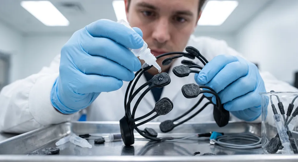
Gamma activity can be measured with several methods, each offering a different balance of timing, spatial information, and susceptibility to artifacts.
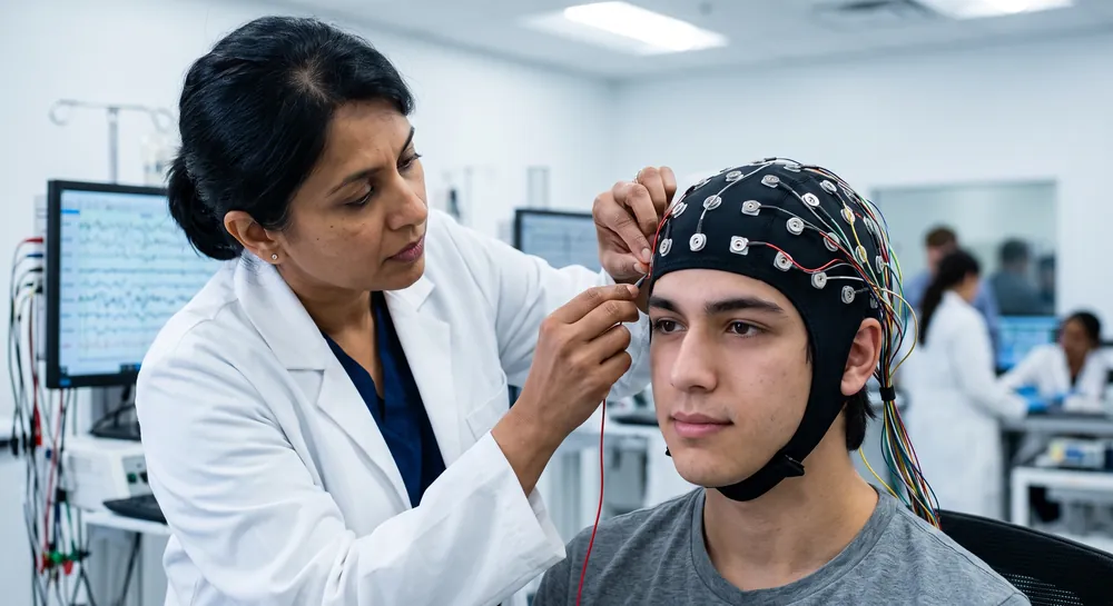
Scalp EEG records voltage changes associated with synchronized neural activity and offers millisecond-level temporal resolution. Magnetoencephalography, or MEG, records magnetic fields generated by neural currents and provides another noninvasive view of oscillatory activity.
Analysis often begins with preprocessing, including removal or marking of movement, eye, muscle, and electrical artifacts. Researchers may then calculate spectral power, phase synchronization, event-related changes, or time-frequency representations. The choice of method can substantially affect whether a brief burst, sustained rhythm, or cross-region relationship is detected.
Interpretation also depends on electrode placement, reference choice, sample size, statistical controls, and the task performed during recording. High-frequency signals at the scalp can be especially sensitive to facial and scalp muscle activity.
A credible conclusion therefore combines signal quality checks with behavioral or clinical data rather than treating a colorful chart as a diagnosis.
Where Gamma Waves Originate in the Brain
Gamma oscillations emerge from the coordinated behavior of local circuits within the cerebral cortex, shaped by both neurons and a class of support cells once thought to have no fast signaling role.
Recordings taken from all layers of the primary visual cortex (V1) in monkeys suggest a laminar organization. Gamma waves were reported to be initiated in input layer 4 and then propagate upward to the superficial layers and downward to the deep layers of cortex. This pattern parallels the flow of sensory information from the thalamus toward higher‑order cortical regions, marking gamma specifically as a feedforward signal—the brain’s way of rapidly pushing novel details forward for further analysis.
This laminar origin tells us that gamma is a local circuit phenomenon that can radiate through the layers of a cortical column. Yet the generation of these fast rhythms depends on a surprising participant.
In hippocampal slice preparations, selectively blocking the release of signaling molecules from astrocytes (star‑shaped glial cells that surround synapses) significantly shortens the duration of chemically induced gamma oscillations. Building on this, a transgenic mouse model with inducible astrocyte dysfunction showed a marked reduction in EEG gamma power in the awake‑behaving brain, even though traditional neuron‑to‑neuron synaptic transmission remained intact.
Astrocytes, it appears, are essential contributors to the rapid network dynamics that sustain gamma.
How Gamma Waves Coordinate Neural Synchrony
The feedforward propagation of gamma waves is not confined to a single region. The aforementioned simultaneous recordings from V1 and a higher visual area, V4, suggest that gamma waves travel in the feedforward direction: when a visual stimulus first hits the cortex, gamma synchrony in V1 precedes and drives gamma in V4.
Causality was established by microstimulating V1 and observing that it directly elicits gamma oscillations in downstream V4. This directional push makes gamma a candidate for the rapid transmission of sensory details from lower to higher processing stages.
But gamma oscillations do not operate in isolation. Cortical processing simultaneously involves multiple frequency bands, raising a question of how spectrally separate streams are integrated.
In a magnetoencephalography study of healthy adults performing continuous mental arithmetic, researchers found robust cross‑frequency phase synchrony among alpha (\~10 Hz), beta (\~20 Hz), and gamma (\~30–40 Hz) oscillations. These tasks—which required holding and summing numbers in working memory—enhanced not only classical within‑band synchrony, but particularly the phase coupling between gamma and alpha bands.
Crucially, when the task became more demanding (increasing the number of items retained), the most prominent boost in synchrony appeared precisely between gamma and alpha. This finding indicates that gamma acts as a key node in a network that ties together slower, more spatially widespread rhythms into a coherent moment of cognition.
The cross‑frequency coordination offers a plausible mechanism for how the brain integrates fast, local feature processing (gamma) with broader, context‑setting activity (alpha).
The Role of Gamma Oscillations in Working and Recognition Memory
Linking gamma synchrony to specific memory systems sharpens the functional picture. The mental arithmetic task described above demonstrated that gamma‑band synchrony, especially when phase‑locked to alpha, strengthened as working memory load increased. The synchronization between gamma and alpha frequencies tracked the cognitive demand of maintaining and manipulating information over seconds.
Evidence from a different memory domain, recognition memory, further supports a causal relationship between gamma power and behavior, and here the astrocyte connection becomes decisive. The same transgenic mice that showed reduced cortical gamma power due to astrocytic tetanus toxin expression also performed poorly in a novel object recognition test, a standard assay for the ability to distinguish familiar from new items.
Notably, other memory capacities—working memory in a maze and fear conditioning—were unchanged. The deficit was specific to recognition memory, and when the toxin expression was suppressed, both gamma power and novel object recognition performance recovered. These interlocking observations point to a potential role for gamma oscillations in the circuitry underlying familiarity detection.
More broadly, a review of oscillatory synchrony in cortical networks catalogs gamma‑band activity among the rhythms associated with perceptual grouping, attention‑dependent stimulus selection, subsystem integration, working memory, and consciousness. When coordination across distant neural populations is required, gamma synchrony seems to consistently appear.
Its presence in tasks ranging from visual binding to mental addition underscores that gamma is not dedicated to a single cognitive domain; rather, it is a flexible temporal tool that the brain deploys whenever rapid integration of information is needed.
Gamma Abnormalities as a Window into Brain Disorders
Because gamma synchrony supports core cognitive operations, alterations in the gamma band can signal underlying pathology.
Schizophrenia provides one of the most studied examples. An event‑related EEG study compared 22 healthy controls and 21 medicated patients during a simple auditory oddball task. Using time‑frequency analysis, researchers measured gamma‑band (36–50 Hz) phase locking—the consistency with which neural responses align in time across trials—within the first 100 milliseconds after a standard tone.
Healthy subjects exhibited prominent phase locking at frontal electrodes between 20 and 60 milliseconds. In the schizophrenia group, this early‑evoked gamma synchrony was significantly reduced (P \= .03), indicating that the brain’s initial synchronization to an auditory stimulus appears to be deficient in the brain disorder.
This finding aligns with a larger body of work suggesting that schizophrenia involves abnormal neural oscillations and synchrony, particularly in the gamma band, and that these abnormalities relate to cognitive dysfunctions and symptoms. Reduced gamma phase locking may represent a pathophysiological mechanism contributing to the fragmentation of thought and perception that characterizes the illness.
While the study underscores the potential of gamma synchrony as a biomarker, it also cautions that methodological choices including the specific time‑frequency decomposition parameters can heavily influence results, and prior studies have not always detected the same deficit. The emerging consensus, however, places gamma‑band synchrony among the most promising electrophysiological leads for understanding and eventually stratifying neuropsychiatric conditions.
Why Gamma Waves Are Central to Brain Coordination and Health
Gamma waves are the brain’s fastest rhythms, acting as a timing signal that pulls separate pieces of information into a coordinated mental experience. Herein we show that these rhythms depend on both neurons and specialized support cells, and that they move forward through the cortex as a way to pass newly arriving sensory details along for further processing. Gamma does not work alone, however; it locks with slower rhythms like alpha when tasks demand more working memory, revealing that flexible cognition rests on precise cross-frequency coordination.
Distortions in this timing may signal disease. When support-cell function was disabled in experiments, gamma power dropped and recognition memory suffered, while people with schizophrenia showed weakened early gamma responses to simple sounds.
These findings support gamma synchrony as a potential marker of how well the brain integrates information, not as a proven cause of any condition. Measuring gamma rhythms in individuals could eventually help clinicians detect and monitor disorders in which the brain’s fastest signaling falls out of step.
References
Van Kerkoerle, T., Self, M. W., Dagnino, B., Gariel-Mathis, M. A., Poort, J., Van Der Togt, C., & Roelfsema, P. R. (2014). Alpha and gamma oscillations characterize feedback and feedforward processing in monkey visual cortex. Proceedings of the national academy of sciences, 111(40), 14332-14341. https://doi.org/10.1073/pnas.1402773111
Lee, H. S., Ghetti, A., Pinto-Duarte, A., Wang, X., Dziewczapolski, G., Galimi, F., Huitron-Resendiz, S., Piña-Crespo, J. C., Roberts, A. J., Verma, I. M., Sejnowski, T. J., & Heinemann, S. F. (2014). Astrocytes contribute to gamma oscillations and recognition memory. Proceedings of the National Academy of Sciences of the United States of America, 111(32), E3343–E3352. https://doi.org/10.1073/pnas.1410893111
Palva, J. M., Palva, S., & Kaila, K. (2005). Phase synchrony among neuronal oscillations in the human cortex. The Journal of Neuroscience, 25(15), 3962-3972. https://doi.org/10.1523/JNEUROSCI.4250-04.2005
Uhlhaas, P. J., Haenschel, C., Nikolić, D., & Singer, W. (2008). The role of oscillations and synchrony in cortical networks and their putative relevance for the pathophysiology of schizophrenia. Schizophrenia bulletin, 34(5), 927-943. https://doi.org/10.1093/schbul/sbn062
Roach, B. J., & Mathalon, D. H. (2008). Event-related EEG time-frequency analysis: an overview of measures and an analysis of early gamma band phase locking in schizophrenia. Schizophrenia bulletin, 34(5), 907-926. https://doi.org/10.1093/schbul/sbn093
Frequently Asked Questions
What are gamma waves and why are they important for brain function?
Gamma waves are the brain's fastest rhythmic electrical activities, measurable using EEG. They enable rapid synchronization of neurons across networks, which helps bind sensory details into unified perceptions and supports cognitive functions like attention, memory, and conscious processing.
Where do gamma waves originate in the brain, and how do they travel?
Gamma waves originate in the input layer (layer 4) of the cerebral cortex and then propagate to superficial and deep layers. This pattern parallels the flow of sensory information, marking gamma as a feedforward signal that rapidly pushes novel details forward for higher-order analysis.
What role do astrocytes play in generating gamma waves?
Astrocytes, star-shaped glial cells surrounding synapses, are essential contributors to gamma oscillations. Blocking their signaling shortens gamma oscillations, and causing their dysfunction reduces gamma power in the brain even when traditional neuron-to-neuron transmission remains intact.
How do gamma waves coordinate with other brain rhythms?
Gamma oscillations integrate with slower rhythms like alpha and beta through cross-frequency phase synchrony. This coordination allows fast, local feature processing in gamma to be tied together with broader, context-setting activity in slower rhythms during tasks like mental arithmetic.
How is gamma activity linked to working memory?
Gamma-band synchrony strengthens as working memory load increases, especially when phase-locked to alpha oscillations. This synchronization tracks the cognitive demand needed to hold and manipulate information over short periods.
What is the connection between gamma power and recognition memory?
Gamma power appears necessary for recognition memory, as reducing it through astrocyte dysfunction selectively impairs novel object recognition. When gamma power is restored, the recognition memory ability recovers, while other memory types remain unaffected.
Why is the feedforward direction of gamma waves significant?
The feedforward propagation from primary visual cortex (V1) to higher visual area (V4) confirms that gamma acts as a rapid transmission mechanism. This directional push supports the quick transfer of sensory details from lower to higher processing stages, enabling coherent perception.
Emotiv is a neurotechnology leader helping advance neuroscience research through accessible EEG and brain data tools.
Medical Disclaimer: The information provided on this website is for educational and informational purposes only and is not intended as medical or health advice. This content may contain errors and should not be relied upon to make life-altering health, medical, or lifestyle choices. Always seek the advice of your physician or other qualified health provider with any questions you may have regarding a medical condition or treatment.
Christian Burgos