Delta waves are slow, high‑amplitude electroencephalogram (EEG) oscillations, typically defined as 0.5 – 4 Hz.
For decades, high‑amplitude delta was almost synonymous with unconsciousness, deep non‑REM sleep, general anesthesia, coma, and the vegetative state. Yet a growing body of research reveals that prominent delta activity can appear in people who are wide awake, responsive, and even experiencing powerful psychedelic states.
What Are Delta Waves?
Delta waves are low-frequency patterns of electrical activity in the brain. They become especially prominent during the deepest stages of non-REM sleep, when awareness of the external environment is reduced and the body enters a highly restorative state.
The phrase “delta waves sleep” therefore refers both to a measurable brain rhythm and to the stage of sleep in which that rhythm is most visible. Noteworthy, delta activity is not, by itself, a complete measure of sleep quality.
Characteristics of Delta Waves
Delta waves have relatively long cycles and low frequency. During slow-wave sleep, they often appear as large, synchronized oscillations across groups of neurons. This synchronization is associated with reduced responsiveness to the environment and with the deepest portion of the normal sleep cycle.
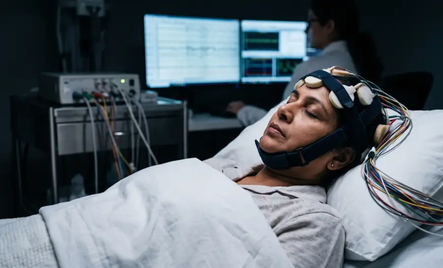
Delta activity is assessed through an EEG, rather than through a consumer sleep score alone. Clinical interpretation considers the full recording, the person’s age and condition, artifacts in the signal, medication or substance exposure, and the relationship between brain activity and other measurements such as breathing and muscle tone. Delta activity during waking can have different meanings from delta activity during sleep, which is why context matters.
Age also influences the amount and distribution of slow-wave activity. Children and adolescents generally show more slow-wave sleep than older adults, while the depth and continuity of this activity may change across the lifespan. These normal differences make broad claims about a “perfect” amount of delta activity unreliable.
How Delta Waves Are Generated in Non‑Sleep States
Delta waves emerge from the cortex itself. Large groups of cortical neurons alternate near‑synchronously between states of hyper‑excitability (“up”) and hypo‑excitability (“down”). The amplitude and slope of a delta wave reflect the number of neurons oscillating together.
Because slower rhythms recruit especially large cell assemblies, the frequency of a delta oscillation is proposed to be inversely related to the size of the synchronized population—a delta rhythm engages a far broader network than a faster alpha or beta rhythm.
This cortical origin explains why delta requires an intact cortex. In stroke models highlighted by Assenza & Lazzaro, a subcortical lesion that removes incoming input triggers delta activity in the overlying cortex, but a lesion that directly damages the cortex to the point of near‑silence does not produce the same delta pattern. In other words, delta arises when cortical circuits are still operational but disconnected from their normal thalamic or subcortical drive.
The relationship between delta and deeper brain structures becomes clearer during propofol‑induced loss of consciousness. According to Boly et al., as propofol deepens, slow activity in the delta‑to‑alpha range rises selectively. When researchers used dynamic causal modeling to trace the source of these spectral changes, they found that the transition to unconsciousness was not driven by a further boost in thalamic excitability; that increase happened earlier during mild sedation.
Instead, loss of consciousness was marked by a specific drop in backward corticocortical connectivity from frontal to parietal areas. So while delta is generated by cortical circuits, what it “means” depends critically on how those circuits talk to each other and to the thalamus.
High‑Amplitude Delta Does Not Automatically Mean “Unconscious”
Clinicians have long used high‑amplitude delta as a red flag for reduced consciousness. In many cases, scuhas slow‑wave sleep, anesthesia, post‑ictal states, it is.
But Frohlich et al. 2021 study documents a striking, under‑acknowledged list of conscious or responsive conditions that also feature prominent delta:
Angelman syndrome
Certain epilepsies
Behavioral responsiveness during propofol anesthesia
Postoperative delirium
States of dissociation such as dreaming
Powerful psychedelic experiences
Older clinical reports add:
Rett syndrome
Lennox‑Gastaut syndrome
Schizophrenia
Mitochondrial diseases
Hepatic encephalopathy
Non‑convulsive status epilepticus.
Delta appears so reliably across this landscape that using delta power alone as a consciousness meter would mislabel many awake, aware individuals as unconscious.
The resolution to this paradox lies in combining spectral power with measures of EEG complexity, or entropy. A delta‑rich EEG can belong to a conscious brain if the signal remains complex and unpredictable; it signals unconsciousness far more reliably when complexity drops.
The propofol data from Boly et al. reinforce this view. Loss of consciousness was not just “more delta,” but a breakdown in frontal‑to‑parietal feedback connectivity, a shift that likely reduces integrated information processing.
Thus, whenever possible, clinicians and researchers should examine connectivity and complexity alongside spectral power.
Awake Delta and Neural Plasticity
Neural plasticity is the brain’s lifelong ability to adjust synaptic strength and reorganize networks. Assenza & Lazzaro explored whether delta waves might serve as a non‑invasive EEG marker of plasticity. They used a technique called transcranial magnetic stimulation (TMS), specifically an intermittent theta‑burst protocol (iTBS), to transiently increase cortical excitability in healthy awake volunteers.
After real iTBS, delta power increased. After sham stimulation, it did not. This temporal coupling suggests an association between plasticity‑like processes and delta, extending the sleep‑plasticity work of Tononi and colleagues into the waking state.
However, the authors explicitly label the data as correlational. No correlation between delta power and motor‑evoked potential amplitude was found, and the delta increase was not cleanly separated in time from the TMS‑induced excitability change. The exact relationship therefore remains unestablished.
Delta Waves in Stroke
Acute stroke offers a natural experiment in which the brain is both injured and simultaneously reorganizing. Assenza & Lazzaro also reviewed evidence showing that delta waves over the affected hemisphere are a sensitive indicator of neuronal dysfunction, correlating with lesion volume and the severity of the acute neurological deficit.
If delta spreads from the affected side to the unaffected hemisphere, prognosis appears to be poorer. Importantly, EEG delta measures in the acute phase add predictive value for clinical recovery at three months, over and above what a clinician can estimate from bedside examination alone.
At the same time, delta may not be purely a sign of damage. In stroke rats, delta in the contralesional hemisphere appears to act as an “attraction guide” for interhemispheric fibers sprouting across to the injured side. The same review hypothesizes that, in human patients, perilesional neurons forging new connections may drive larger assemblies to oscillate at delta frequencies, reflecting an attempted neural rearrangement.
So the delta seen after stroke likely embodies both initial injury and ongoing, albeit incomplete, recovery processes. Again, the evidence is correlational; no causal role for delta in shaping plasticity or repair has been proven.
Non‑Sleep Delta and Brain Clearance
The glymphatic system is a network that moves cerebrospinal fluid (CSF) alongside perivascular spaces and through brain tissue, delivering nutrients and clearing metabolic waste.
A landmark mouse study by Hablitz et al. examined glymphatic influx under six different anesthesia regimens and compared it with brain activity and cardiovascular measures. CSF tracer influx was highest under ketamine‑xylazine, followed by isoflurane supplemented with dexmedetomidine and pentobarbital; it was low under α‑chloralose, Avertin, and isoflurane alone.
When the researchers looked across all regimens, they found a positive correlation between glymphatic influx and cortical delta power, and a negative correlation with beta power and heart rate. This is a striking observation, but it records an association in anesthetized animals, not a causal mechanism in awake humans.
Extrapolations that “delta waves cleanse the brain” or that waking delta acutely boosts glymphatic flow oversimplify the data and are not supported for non‑sleep states by the evidence discussed.
What Delta Waves Reveal About Brain Function
Delta waves can appear in awake and responsive brains, including psychedelic states, and their meaning depends on the brain's overall connectivity and signal complexity rather than amplitude alone. That shift matters for anyone interpreting EEG evidence in clinics or research settings.
The studies reviewed also link awake delta to plasticity and stroke recovery, but only as an association, not a proven cause. A similar caution applies to links between delta and brain-clearing fluid flow, which come from anesthetized mice and do not prove an effect in humans.
The practical lesson is to combine spectral power with complexity and connectivity measures before drawing conclusions about consciousness, injury, or healing.
References
Assenza, G., & Di Lazzaro, V. (2015). A useful electroencephalography (EEG) marker of brain plasticity: delta waves. Neural regeneration research, 10(8), 1216. https://doi.org/10.4103/1673-5374.162698
Boly, M., Moran, R., Murphy, M., Boveroux, P., Bruno, M. A., Noirhomme, Q., ... & Friston, K. (2012). Connectivity changes underlying spectral EEG changes during propofol-induced loss of consciousness. The Journal of Neuroscience, 32(20), 7082-7090. https://doi.org/10.1523/JNEUROSCI.3769-11.2012
Frohlich, J., Toker, D., & Monti, M. M. (2021). Consciousness among delta waves: a paradox?. Brain, 144(8), 2257-2277. https://doi.org/10.1093/brain/awab095
Hablitz, L. M., Vinitsky, H. S., Sun, Q., Stæger, F. F., Sigurdsson, B., Mortensen, K. N., ... & Nedergaard, M. (2019). Increased glymphatic influx is correlated with high EEG delta power and low heart rate in mice under anesthesia. Science advances, 5(2), eaav5447. https://doi.org/10.1126/sciadv.aav5447
Frequently Asked Questions
What are delta waves?
Delta waves are slow, high-amplitude brain oscillations measured by EEG, typically falling in the 0.5 to 4 Hz range. They reflect rhythmic neural firing patterns where large groups of cortical neurons synchronize between hyper-excitable and hypo-excitable states.
Why were delta waves historically linked to unconsciousness?
High-amplitude delta was considered nearly synonymous with unconsciousness because it appears during deep non-REM sleep, general anesthesia, and coma. This was thought to reflect widespread cortical deactivation during "down states," when large sections of the cortex fall nearly silent.
Can delta waves appear in awake, conscious people?
Yes, prominent delta activity has been documented in awake and responsive individuals, including those in psychedelic states, certain epilepsies, and dream-like dissociative states. Using delta power alone as a consciousness meter could mislabel alert people as unconscious.
How can clinicians distinguish between delta that signals unconsciousness and delta in awake states?
The key is to pair spectral power with measures of EEG complexity or entropy. Delta-rich EEG can belong to conscious brains when the signal remains complex and unpredictable, while unconsciousness is more reliably signaled when complexity drops.
Do delta waves prove that brain plasticity is happening?
Delta activity is associated with plasticity-like processes, as seen after transcranial magnetic stimulation in awake adults, but this evidence is correlational. No causal role has been proven, so delta as a plasticity marker is an exciting hypothesis, not a proven fact.
Do delta waves cleanse the brain by boosting glymphatic flow?
A mouse study found a positive correlation between delta power and glymphatic influx under anesthesia, but this is an association in anesthetized animals, not causal evidence in awake humans. Claims that "delta cleanses the brain" in waking states oversimplify the data.
What should clinicians consider before interpreting delta waves?
Delta should never be interpreted in isolation; spectral power should be combined with measures of connectivity and complexity. This approach prevents misjudging conscious individuals as unconscious based on delta amplitude alone.
Emotiv is a neurotechnology leader helping advance neuroscience research through accessible EEG and brain data tools.
Medical Disclaimer: The information provided on this website is for educational and informational purposes only and is not intended as medical or health advice. This content may contain errors and should not be relied upon to make life-altering health, medical, or lifestyle choices. Always seek the advice of your physician or other qualified health provider with any questions you may have regarding a medical condition or treatment.
Christian Burgos


