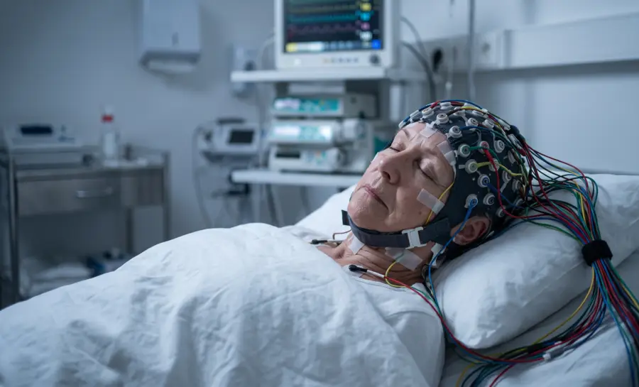
Sleep is often described as a quiet retreat from sensory life, but at the level of brain waves it is a highly active and precisely sequenced state.
Electroencephalography (EEG) records this activity from the scalp, and modern neuroscience uses those recordings to show that the sleeping brain moves through an orderly progression of oscillatory modes, from lighter non-REM (NREM) sleep into deep slow-wave sleep and then into rapid eye movement (REM) sleep. Each mode has its own electrical signature, and each signature reflects different patterns of cellular and network activity.
Brain Wave Patterns During Sleep Stages
Sleep is organized into repeating cycles rather than a single uniform condition. Non-REM sleep progresses from light sleep into deeper slow-wave sleep, followed by periods of REM sleep that become longer later in the night. Brain waves help distinguish these stages, but sleep staging also considers eye movements, muscle tone, breathing, and other signals.
Sleep stage | Typical EEG features | General role in sleep architecture |
|---|---|---|
N1 | Reduced alpha and increasing theta | Transition into sleep |
N2 | Theta background, spindles, and K-complexes | Stable non-REM sleep and preparation for deeper sleep |
N3 | High-amplitude, slow delta activity | Deep slow-wave sleep |
REM | Low-amplitude, mixed-frequency activity | Dreaming, memory, and emotional processing contexts |
Stage N1: Alpha Brain Waves for Sleep Onset
Stage N1 is the brief transition from wakefulness to sleep. Alpha activity, which is typical of relaxed wakefulness, gradually diminishes as slower theta activity becomes more prominent. Muscle activity begins to relax, awareness of the environment fades, and people may experience drifting thoughts or brief sensations of falling.
N1 is not usually considered restorative deep sleep. It is a threshold state, and brief arousals can return the brain to wakefulness. The presence of alpha activity near sleep onset therefore does not mean that the brain has entered a stable or deep sleep stage.
Stage N2: Best Brain Waves for Deep Sleep
Stage N2 is still light-to-moderate non-REM sleep, although it generally occupies a substantial portion of the night. EEG recordings show a background of theta activity interrupted by sleep spindles and K-complexes, two characteristic features used in formal sleep staging. These events reflect organized changes in brain activity rather than a single continuous rhythm.
Stage N3: Deep Sleep (Slow-Wave Sleep)
Stage N3 is the deepest non-REM stage and is characterized by prominent, high-amplitude slow waves, usually described as delta activity. It is more difficult to awaken from N3 than from lighter sleep, and this stage is concentrated earlier in the biological night for many people. The amount and expression of slow-wave sleep can vary with age, prior sleep, circadian timing, and health.
REM Sleep: The Dreaming Stage
REM sleep is associated with vivid dreaming for many people, rapid eye movements, and marked reduction of skeletal muscle tone. Its EEG pattern is generally low-amplitude and mixed-frequency, making it more similar to wakefulness than to N3 sleep. Theta activity and other rhythms may appear, but REM is identified through the combined pattern of brain, eye, and muscle signals.
How the Brain Shifts From Wakefulness to Sleep
The transition from wakefulness to sleep is a coordinated shift in the way the cortex and thalamus communicate, a switch in the brain's default operating mode. The network that connects these regions, called the thalamocortical system, produces much of the rhythmic electrical activity recorded at the scalp.
During wakefulness, neurons in this system fire irregularly and respond selectively to incoming sensory information. When sleep begins, the same network shifts into a highly synchronized pattern dominated by slow oscillations of membrane potential, the electrical voltage across each neuron's membrane.
In this rhythm a cortical neuron alternates between a hyperpolarized down-state, when it is nearly silent, and a depolarized up-state, when it fires intensely. The cycle repeats at a frequency below 1 Hz.
Modeling the Wake-to-Sleep Neural Transition
Understanding this transition requires moving from single neurons to large networks. A large-scale computer model, built from thousands of neurons with several intrinsic currents and millions of interconnections across visual cortex and thalamic nuclei, reproduced both wake and sleep activity patterns.
In the waking mode, the model shows irregular spontaneous firing and selective responses to visual input. In the sleep mode, neuromodulatory changes push the network into slow oscillations that closely resemble those recorded in living tissue.
Systematic exploration of this model showed that an increase in potassium leak conductances is sufficient to trigger the transition from wakefulness to sleep. Once the network enters the sleep mode, the activation of persistent sodium currents initiates the up-state.
A combination of intrinsic and synaptic currents then maintains that up-state, while depolarization-activated potassium currents and synaptic depression bring it to an end. Corticocortical connections then synchronize the slow oscillation across the cortex. This cellular rhythm is a basic engine of sleeping brain activity, an engine that organizes other brain waves through the night.
Traveling Slow Waves: A Spatiotemporal Blueprint for Sleep
The slow oscillation may begin as a cellular event, but it does not remain local. Using high-density EEG, researchers Massimini et al. showed that each cycle of the slow oscillation is a traveling wave. Rather than activating the whole cortex at once, the wave originates at a definite site and moves across the scalp at an estimated speed of 1.2 to 7.0 meters per second.
Most waves arise in anterior regions, often in prefrontal and orbitofrontal areas just behind the forehead, and propagate toward the back of the brain in an anteroposterior direction. As sleep deepens, these waves occur more frequently, eventually reaching almost once per second. This accelerating rhythm begins to align with the high-amplitude delta waves that characterize the deepest stages of NREM sleep.
What makes this spatial pattern significant is its repeatability. The origin sites and propagation routes of slow oscillations are reproducible across different nights and even across different people. That consistency suggests the traveling wave behaves like a map of individual cortical connectivity and excitability, reflecting the underlying wiring of each sleeping brain.
Moreover, the same coordinated wave may also play a learning role. Spike timing-dependent synaptic plasticity is a neural mechanism in which the precise timing between firing events determines whether a synapse strengthens or weakens.
When a wave of correlated activity travels through connected neurons, it can create the timing relationships that drive this form of plasticity during sleep. Therefore, the traveling slow oscillation is suggested to supply a spatial and temporal framework for reorganizing cortical connections across the night.
How Spindles and Ripples Consolidate Memory
One of the principal functions of slow oscillation waves appears to be organizing faster sleep rhythms, particularly sleep spindles and sharp-wave ripples.
Sleep spindles are brief bursts of oscillatory activity in a distinct frequency band, and sharp-wave ripples are even faster events, brief oscillations near 200 Hz produced by tightly synchronized firing in the hippocampus. The slow oscillation provides a repeating temporal structure, and within that structure the faster events find their timing.
When these faster rhythms become nested within particular phases of the slower oscillation, the brain is performing a form of cross-frequency coupling, a timing relationship in which slower rhythms impose order on faster ones. This coupling is considered central to how sleep supports memory.
Direct evidence comes from recordings of hippocampal neurons during slow wave sleep in rats. The hippocampus plays a key role in converting experience into stable neural traces, and during slow wave sleep it spontaneously reactivates activity patterns from prior waking behavior.
One way to measure this reactivation is to track pairwise firing-rate correlations, the tendency of pairs of CA1 pyramidal cells to fire together. The distribution of these correlations during a familiar experience explains a significant portion of the variance in firing patterns during subsequent slow wave sleep and quiet wakefulness.
The strongest reinstatement was reported to occur during sharp wave-ripple oscillations. That timing suggests ripples may be moments when the hippocampal network converges onto attractor states, stable activity patterns that correspond to previous experiences.
Why the Night’s Brain Rhythms Matter for Memory and Arousal
Sleep is an active, highly structured neurological process driven by a precise sequence of electrical rhythms. The seamless transition from wakeful thalamocortical firing to synchronized, slow oscillations below 1 Hz initiates a global shift that coordinates the entire cortex.
As these traveling slow waves propagate along consistent paths, they map individual neural connectivity and set a spatiotemporal stage for synaptic plasticity. Within this slow-wave framework, the nesting of faster sleep spindles and hippocampal sharp-wave ripples—known as cross-frequency coupling—allows the brain to replay and solidify memories of waking experiences.
Ultimately, understanding these intricate, synchronized oscillations reveals how sleep acts as a critical engine for neural reorganization, memory consolidation, and overall cognitive maintenance.
References
Hill, S., & Tononi, G. (2005). Modeling sleep and wakefulness in the thalamocortical system. Journal of neurophysiology, 93(3), 1671-1698. https://doi.org/10.1152/jn.00915.2004
Massimini, M., Huber, R., Ferrarelli, F., Hill, S., & Tononi, G. (2004). The sleep slow oscillation as a traveling wave. The Journal of Neuroscience, 24(31), 6862-6870. https://doi.org/10.1523/JNEUROSCI.1318-04.2004
Kudrimoti, H. S., Barnes, C. A., & McNaughton, B. L. (1999). Reactivation of hippocampal cell assemblies: effects of behavioral state, experience, and EEG dynamics. The Journal of Neuroscience, 19(10), 4090-4101. https://doi.org/10.1523/JNEUROSCI.19-10-04090.1999
Prerau, M. J., Brown, R. E., Bianchi, M. T., Ellenbogen, J. M., & Purdon, P. L. (2017). Sleep neurophysiological dynamics through the lens of multitaper spectral analysis. Physiology, 32(1), 60-92. https://doi.org/10.1152/physiol.00062.2015
Frequently Asked Questions
What happens in the brain when we fall asleep?
The transition from wakefulness to sleep is a coordinated shift in how the cortex and thalamus—the thalamocortical system—communicate. Neurons change from firing irregularly in response to sensory input to firing in a highly synchronized pattern dominated by slow oscillations.
What is the slow oscillation?
The slow oscillation is a rhythm where a cortical neuron alternates between a nearly silent hyperpolarized down-state and an active depolarized up-state, repeating at a frequency below 1 Hz. It acts as a basic engine that organizes other brain rhythms during the night.
What are traveling slow waves?
Each cycle of the slow oscillation is a traveling wave that originates at a specific site, usually in the anterior regions of the brain, and propagates toward the back. The origin sites and propagation routes are reproducible across nights and people, suggesting they reflect the brain's underlying wiring and connectivity.
How does sleep help consolidate memory?
During slow wave sleep, the hippocampus reactivates patterns of activity from prior waking experiences, with the strongest reactivation occurring during sharp-wave ripples. The slow oscillation organizes faster rhythms like spindles and ripples, and this coupling is central to memory consolidation.
Emotiv is a neurotechnology leader helping advance neuroscience research through accessible EEG and brain data tools.
Medical Disclaimer: The information provided on this website is for educational and informational purposes only and is not intended as medical or health advice. This content may contain errors and should not be relied upon to make life-altering health, medical, or lifestyle choices. Always seek the advice of your physician or other qualified health provider with any questions you may have regarding a medical condition or treatment.
Christian Burgos