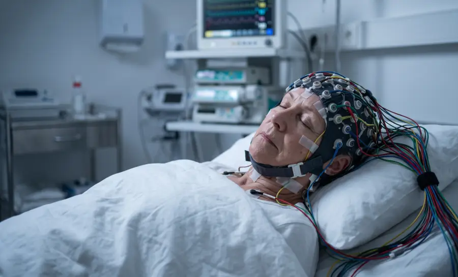
Deep non-rapid eye movement (NREM) sleep narrows into a distinct stage called slow-wave sleep (SWS), or stage N3. On an electroencephalogram (EEG), this stage is defined by high-amplitude, low-frequency electrical activity.
The term "delta waves" can refer to one specific voltage band, but in sleep research it often works more broadly. It covers delta waves measured between 75 and 140 microvolts, EEG slow waves above 140 microvolts, the slow oscillation below 1 Hz, and slow-wave activity in the 0.75 to 4.5 Hz band.
The Role of Delta Waves for Sleep and Rest
Delta waves are closely linked with the restorative part of sleep, but they do not act as a separate treatment or a switch that can simply be turned on. They emerge as part of coordinated changes in the brain, body temperature, hormones, autonomic activity, and sleep pressure.
The timing of these changes is governed by both the circadian rhythm and the need for recovery accumulated during wakefulness. A night with sufficient total sleep can still feel unrefreshing if it is fragmented or poorly aligned with biological timing.
How Cortical Slow Oscillations Generate Traveling Delta Waves
The electrical signature of N3 begins at the level of cortical neurons. From a neuroscience perspective, the fundamental event is the slow oscillation below 1 Hz.
Each cycle alternates between two states:
The first is a hyperpolarized down state, a phase in which the neuron's membrane potential moves further from the firing threshold and activity quiets.
The second is a depolarized up state, a phase in which the membrane potential shifts closer to the firing threshold and cells fire intensely.
This alternation synchronizes across large neuronal populations and also organizes other sleep rhythms, including spindles and slow waves.
High-density EEG recordings reveal that each cycle of this slow oscillation behaves as a traveling wave. It starts at a definite site, most often in prefrontal-orbitofrontal regions, and then moves over the scalp in an anteroposterior direction. Further, the estimated speed is between 1.2 and 7.0 meters per second.
As sleep deepens, the rate of these waves increases, approaching almost once per second. This pattern is often part of the broader normal adult EEG waveform profile across sleep stages.
Research suggests that the origin and propagation pattern is reproducible across nights and across subjects. Researchers describe it as a blueprint of cortical excitability and connectivity.
A wave tends to follow the same general route because that route reflects stable properties of the underlying cortex. The orderly propagation of correlated activity along connected pathways may contribute to spike timing-dependent synaptic plasticity, a process in which the relative timing of neuronal firing influences connection strength. This mechanism becomes relevant again when considering memory consolidation.
The active nature of this state appears with simultaneous EEG and functional MRI. In 14 non-sleep-deprived healthy volunteers, slow waves above 140 microvolts and delta waves between 75 and 140 microvolts were linked to significant increases in brain activity in several cortical areas. The involved regions included the inferior frontal, medial prefrontal, precuneus, and posterior cingulate areas.
Slow waves also engaged the parahippocampal gyrus, cerebellum, and brainstem. Additionally, delta waves showed a more frontal response.
That evidence directly challenges the older view of SWS as a period of brain quiescence. During N3, specific cerebral regions synchronize to the slow oscillation and become active.
Why Delta Activity Tracks Homeostatic Sleep Pressure
Delta activity also tracks how much sleep pressure has accumulated. Slow-wave activity, the EEG power in the slow and delta range, decreases over the course of a sleep episode. This decline happens even when the timing of sleep is experimentally moved.
In one protocol, eight men lived in an environment free of time cues for 33 to 36 days and followed a 28-hour rest-activity cycle. Sleep episodes lasted 9.33 hours and occurred at all phases of the endogenous circadian cycle. The design separated circadian timing from homeostatic sleep pressure.
Under these conditions, slow-wave activity in non-REM sleep decreased during every sleep episode. That pattern identifies slow-wave activity as an index of the sleep homeostat, the internal process that tracks sleep need.
Slow-wave activity also showed a low-amplitude circadian modulation, but that modulation did not parallel the circadian rhythm of sleep propensity. The same study found a strong circadian rhythm in REM sleep, with its crest positioned shortly after the minimum of the core body temperature rhythm.
Moreover, sleep spindle activity showed a marked endogenous circadian rhythm, with its crest near habitual sleep onset. Yet, slow-wave activity did not follow those rhythms. This contrast supports the idea that delta activity primarily reflects homeostatic sleep pressure rather than circadian clock output.
Experimental suppression makes the homeostatic link even clearer. Tasali et al. showed that in healthy young adults, all-night selective suppression of SWS reduced delta spectral power without changing total sleep time. The intervention reduced the dominant EEG frequency range of SWS and left other EEG frequency bands unchanged.
Delta activity can therefore be suppressed independently of sleep duration. That same experimental design also opened a direct test of whether SWS matters for the body outside the brain.
How Slow Oscillations Couple the Neocortex and Hippocampus
The functional value of these oscillations appears partly in their timing relationship with hippocampal activity. Slow oscillations originating in the prefrontal neocortex group neuronal network activity during N3. This grouping influence extends to hippocampal sharp wave-ripple activity.
Sharp wave-ripples are brief, high-frequency hippocampal events thought to support the replay of waking experience during sleep. In rats, Mölle et al. recorded prefrontal surface EEG, prefrontal multiunit activity, and hippocampal local field potentials from CA1 during sleep. They calculated ripple and spindle activity time-locked to the half-waves of slow oscillations.
Therein, they found that ripple activity and sharp wave-ripples decreased during the negative half-wave of the slow oscillation and increased during the positive half-wave. The depolarizing surface-positive phase, and the associated up state of prefrontal excitation, appears to promote hippocampal sharp wave-ripples through efferent pathways.
Fine-grained analysis showed that up and down states in prefrontal multiunit activity preceded corresponding changes in hippocampal sharp wave-ripples by roughly 30 milliseconds. This timing indicates that the neocortex may drive the temporal coordination rather than simply following hippocampal output.
Moreover, the traveling wave properties described earlier add a spatial dimension to this idea. The orderly propagation of slow oscillations along connected pathways may contribute to spike timing-dependent synaptic plasticity during sleep.
When a wave passes through connected regions in a reliable sequence, the timing of neuronal firing becomes reproducible. That reproducibility is a candidate mechanism for memory consolidation, because the precise order of firing can influence which connections strengthen.
Human neuroimaging data offer another clue. The brain response pattern associated with SWS waves partially overlaps with the waking default mode network, a set of regions active during wakeful rest and internally directed thought.
This overlap is consistent with the hypothesis that slow oscillations restore microwake-like activity patterns that facilitate neuronal interactions. The phrase microwake-like does not mean SWS is wakefulness, rather it means that organized patterns of regional communication can reactivate during deep sleep, potentially supporting off-line processing.
Why Slow-Wave Sleep Is a Dynamic Bridge Between Brain and Body
Slow-wave sleep is far from a passive brain state; its delta waves begin as cortical oscillations, travel in repeatable patterns, and reflect accumulated sleep pressure rather than the body clock. These waves time communication between the neocortex and hippocampus, linking deep sleep to memory-related replay processes.
Still, the story is incomplete. Researchers have not yet shown how traveling waves translate into lasting memory gains, how cortical slow oscillations affect glucose regulation, or whether boosting this activity could improve health.
References
Dang-Vu, T. T., Schabus, M., Desseilles, M., Albouy, G., Boly, M., Darsaud, A., ... & Maquet, P. (2008). Spontaneous neural activity during human slow wave sleep. Proceedings of the National Academy of Sciences, 105(39), 15160-15165. https://doi.org/10.1073/pnas.0801819105
Dijk, D. J., & Czeisler, C. A. (1995). Contribution of the circadian pacemaker and the sleep homeostat to sleep propensity, sleep structure, electroencephalographic slow waves, and sleep spindle activity in humans. The Journal of neuroscience, 15(5), 3526-3538. https://doi.org/10.1523/JNEUROSCI.15-05-03526.1995
Tasali, E., Leproult, R., Ehrmann, D. A., & Van Cauter, E. (2008). Slow-wave sleep and the risk of type 2 diabetes in humans. Proceedings of the National Academy of Sciences, 105(3), 1044-1049. https://doi.org/10.1073/pnas.0706446105
Mölle, M., Yeshenko, O., Marshall, L., Sara, S. J., & Born, J. (2006). Hippocampal sharp wave-ripples linked to slow oscillations in rat slow-wave sleep. Journal of neurophysiology. https://doi.org/10.1152/jn.00014.2006
Frequently Asked Questions
What are delta waves and how do they relate to slow-wave sleep?
Delta waves are low-frequency, high-amplitude brain waves that dominate slow-wave sleep (stage N3). In sleep research, the term covers a broad range of slow patterns, including EEG slow waves and slow oscillations below 1 Hz, all of which share the feature of dominating deep NREM sleep.
Are slow-wave sleep and the brain inactive during N3?
No, N3 is not a state of brain silence. Simultaneous EEG and functional MRI recordings show that slow waves and delta waves are linked to synchronized increases in brain activity in several cortical areas, directly challenging the older view of deep sleep as a period of brain quiescence.
Where do slow waves originate and how do they travel across the brain?
Each slow oscillation cycle begins at a definite site, most often in prefrontal-orbitofrontal regions, and then moves over the scalp in an anteroposterior direction as a traveling wave. The propagation pattern is reproducible across nights and subjects, reflecting stable properties of the underlying cortex and its connectivity.
What is the relationship between delta activity and sleep pressure?
Slow-wave activity decreases over the course of a sleep episode even when sleep timing is experimentally shifted, identifying it as an index of homeostatic sleep pressure. Unlike REM sleep and spindle activity, slow-wave activity does not follow circadian rhythms, supporting the idea that delta activity primarily reflects how much sleep need has accumulated.
How do slow oscillations coordinate communication between the neocortex and hippocampus?
Slow oscillations from the prefrontal neocortex group hippocampal sharp wave-ripple activity, which is thought to support memory replay. Ripple activity increases during the positive half-wave of the slow oscillation, and the neocortex drives this timing coordination, as prefrontal changes precede hippocampal changes by roughly 30 milliseconds.
Emotiv is a neurotechnology leader helping advance neuroscience research through accessible EEG and brain data tools.
Medical Disclaimer: The information provided on this website is for educational and informational purposes only and is not intended as medical or health advice. This content may contain errors and should not be relied upon to make life-altering health, medical, or lifestyle choices. Always seek the advice of your physician or other qualified health provider with any questions you may have regarding a medical condition or treatment.
Christian Burgos


