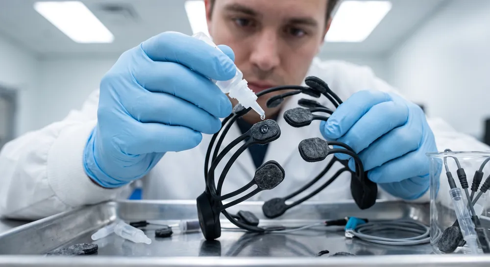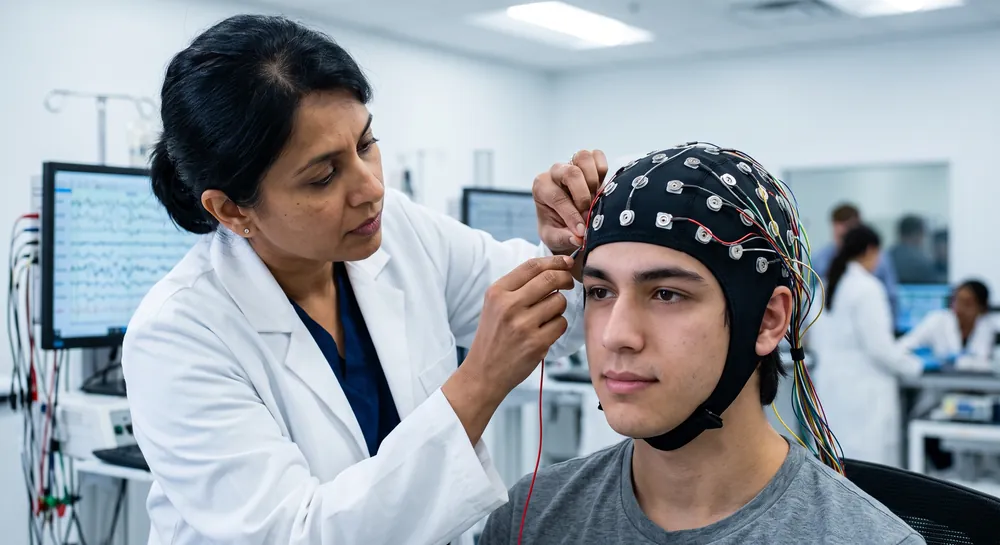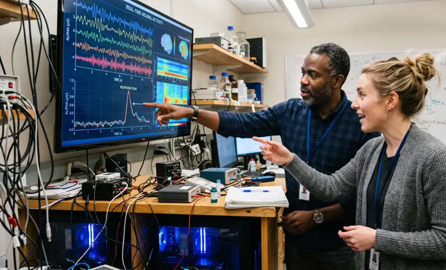
Inside the dark vault of the skull, an unceasing electrical conversation runs at all hours, whether you are solving a complex equation, staring blankly at a wall, or deep in dreamless sleep. This electrical activity, when recorded from the scalp, appears as rhythmic, oscillating waves. These brain waves represent the synchronized activity of vast neural populations and provide one of the few direct windows into the living, functioning human brain.
This overview will explore the physiological origin of these rhythms, the fundamentals of how they are recorded, and their spatiotemporal organization.
What Are Brain Waves?
Brain waves are rhythmic or repeating patterns of electrical activity produced by networks of neurons. They arise as groups of nerve cells communicate and become temporarily synchronized, creating signals that can be detected outside the skull. The term is commonly used in both clinical medicine and neuroscience, although the rhythms themselves are only one part of the brain’s highly complex activity.
The frequency of a brain wave describes how many cycles occur per second, measured in hertz (Hz). Faster rhythms tend to have higher frequencies, while slower rhythms have lower frequencies. These patterns are not fixed labels for permanent personality traits or diagnoses; they change with arousal, sensory input, sleep stage, attention, medication, illness, and the recording conditions.
What EEG Actually Measures at the Scalp
To understand brain waves, one must first appreciate what an EEG electrode genuinely detects. The brain is a highly folded mass of tissue with billions of neurons. When a single neuron fires, the resulting electrical field is minuscule and spatially chaotic.
For a rhythm to appear on an EEG recording, a massive number of neurons must be active in sync. They need to be arranged in a structured, parallel geometry and receive similar inputs at roughly the same time.
The cerebral cortex’s pyramidal neurons are perfectly suited for this since they are aligned perpendicular to the brain’s surface, acting like tiny dipoles. When they receive synchronous input, their summed electrical potential is what propagates through the various layers of tissue and skull to reach a scalp electrode.
However, the path from neuron to scalp is not a simple wire transfer. The EEG signal is a volume-conducted mixture. By the time a signal reaches an electrode, it contains contributions from multiple distinct brain sources, all smeared together through the conductive medium of the head.
Decomposing this mixture remains a core signal processing challenge. Research into independent component analysis algorithms has provided a critical insight into the nature of these hidden sources.
A key finding by Delorme et al. from a comparison of 22 EEG decomposition algorithms revealed that the most effective methods identified sources that were remarkably “dipolar.” This means the scalp map of an isolated source could be modeled as if it were projected by a single, compact equivalent dipole within the brain.
The study concluded that this result is compatible with the interpretation that many independent EEG components are volume-conducted projections of partially-synchronous local field activity within a single, compact cortical domain. In plain language, the rhythmic waves we see on an EEG are not reflecting diffuse, disorganized activity across the whole brain. Instead, they capture the synchronized electrical “hum” of a specific, localized chunk of cortex.
A high-quality EEG system, combined with advanced signal processing, effectively disentangles this mixture to reveal the distinct neural engines working in parallel.
How Neurons Build a Rhythm
The neural architecture generating brain waves arises from the intricate wiring between the cortex and deeper brain structures, particularly the thalamus. Interactions between excitatory and inhibitory neurons form circuits that establish the rhythm within a local patch of tissue.
These circuits can then synchronize with other local patches, creating larger-scale waves. The cellular basis for both very slow and fast rhythms has been modeled in detail.
The Cellular Basis of Slow Oscillation Synchronization
The slowest brain rhythms, those below 1 Hz and characteristic of deep sleep, offer a clear view of synchronized inhibition in action.
An investigation by Contreras & Steriade into the dynamic relationship between the cortex and thalamus during this slow oscillation showed a highly organized ballet of firing and silence. During the depth-positive phase of a slow EEG wave, both cortical neurons and neurons in the reticular thalamic nucleus became hyperpolarized.
In this hyperpolarized state, the cell membrane is held down, making the neuron deeply silent. As the wave shifted to its depth-negative phase, these neurons depolarized, waking up to fire, often in bursts.
Further, during the depth-negative EEG wave, when cortical cells depolarized, thalamocortical neurons received a pattern of inhibitory postsynaptic potentials. The researchers concluded that this specific phase-locking across cortical and thalamic circuits appears to reflect a generalized inhibitory phenomenon. The rhythm of the slow oscillation is fundamentally a rhythm of periods of active inhibition sculpting periods of allowable firing across the entire corticothalamic loop.
This active inhibition pattern must then be a core mechanism for synchronizing vast territories of the brain.
How Fast Oscillations Emerge from Inhibition
The same principle of inhibition-driven rhythm also applies to fast oscillations in the gamma range (20-80 Hz), but through a different circuit.
A computer model of a hippocampal interneuronal network tested how a specific type of neuron, GABAergic fast-spiking interneurons, could generate fast rhythms. These cells, which produce the inhibitory neurotransmitter GABA, were connected in a random network. The simulation found that rhythmic network synchronization could emerge entirely from GABAA synaptic transmission.
The study identified a critical condition for this synchronization to occur across a large network. There had to be a critical minimal average number of synaptic contacts per cell. Once this minimum connectivity threshold was crossed, the network synchronized into a unified rhythm.
Importantly, the model showed high network synchronization only within a limited frequency band that matched the gamma range. The conclusion is that the very mechanism of fast synaptic inhibition, when densely connected, provides a precise temporal framework for synchronization.
One cluster of inhibitory interneurons can rhythmically silence surrounding excitatory neurons, creating the narrow time windows in which they can fire, and thereby orchestrating a high-frequency beat that can synchronize discharges of spatially distributed neurons.
The Spatiotemporal Organization of Brain Waves
Brain waves are spatiotemporally organized events that travel across the cortex in complex ways. This organized movement provides a potential blueprint of the brain’s underlying connectivity.
A high-density EEG study looking at the slow oscillation of sleep demonstrated this directly. Each cycle of the slow oscillation, the large wave of firing and silence, was found to be a traveling wave. An individual wave originates at a definite site on the scalp and then travels over the cortical surface at a speed of 1.2 to 7.0 meters per second.
These waves did not originate randomly. Rather, they were more frequently observed starting in prefrontal and orbitofrontal regions, propagating predominantly in an anterior-to-posterior direction. As a person drifted deeper into slow-wave sleep, the occurrence of these traveling waves increased progressively, reaching a rate of almost one per second.
The pattern of where a wave originates and the path it takes is remarkably consistent. It is reproducible across multiple nights in the same person and is similar across different subjects.
The study’s authors proposed that this ordered propagation of correlated activity along connected pathways could play a role in synaptic plasticity during sleep. The physical path the wave takes is, in effect, retracing the anatomical highways linking these brain regions.
Beyond the spatial journey of a single cycle, brain oscillations also exhibit complex, long-range temporal organization. Fluctuations in the amplitude of a particular rhythm are not random over time.
An investigation using simultaneous magnetoencephalography (MEG) and EEG found that amplitude fluctuations of 10 and 20 Hz oscillations are correlated over incredibly long time scales, spanning thousands of individual oscillation cycles and lasting minutes. These amplitude fluctuations followed a power-law scaling behavior. This means that the pattern of fluctuations looked self-similar whether you examined a short window of seconds or a long window of minutes. The scaling exponents were highly invariant across subjects, indicating a fundamental physiological property.
The researchers proposed that this long-range correlation and power-law scaling finds a unifying explanation in the theory of self-organized criticality. In this view, a neural network naturally tunes itself to a critical state, a point of optimal dynamic range, where it can quickly reorganize its activity in response to processing demands.
Broad Classification of Brain Waves
For clinical and research utility, the continuous spectrum of brain waves is conventionally divided into distinct frequency bands. This classification is descriptive, a set of labels for the dominant rhythmic engine running at a given time, and does not imply that a single frequency is doing a singular, isolated job.
The main bands, from slowest to fastest, are delta, theta, alpha, beta, and gamma.
Brain-wave band | Approximate frequency | Common associations |
|---|---|---|
Delta | 0.5–4 Hz | Deep sleep and slow-wave activity |
Theta | 4–8 Hz | Drowsiness, dreaming, and memory-related activity |
Alpha | 8–13 Hz | Relaxed wakefulness, especially with reduced sensory engagement |
Beta | 13–30 Hz | Alertness, active thinking, and sustained mental effort |
Gamma | About 30 Hz and above | High-frequency integration and complex information processing |
These bands provide a relatively universal vocabulary for describing the state of the cortex. A state dominated by delta waves is qualitatively different from one dominated by beta waves, much in the way that a slow, rolling swell on the ocean is different from a surface covered in small, choppy ripples.
However, a strict definition is that these band labels describe the overall range of rhythmic electrical activity. This overview intentionally refrains from assigning a fixed cognitive or clinical function to any single band.
A gamma oscillation identified in a hippocampal network model might share a frequency with a gamma rhythm in the visual cortex but arise from a different circuit and serve a different computational purpose.
Limitations and Public Perception of Brain Waves
The language of brain waves has become deeply embedded in popular culture. Claims about “entraining alpha waves” for relaxation, “increasing gamma waves” for peak focus, or “boosting theta waves” for creativity are widespread.
It is important to directly address the scope of the evidence presented here. The studies above provide foundational neurophysiological mechanisms. They show that large-scale synchronization is an inhibition-based circuit property. They show that waves travel in organized patterns. They show that oscillations display complex long-memory dynamics.
However, these foundational mechanisms do not directly test most popular commercial claims about brain-wave enhancement. The finding that a computer model of interneurons can generate a gamma rhythm through inhibition does not constitute evidence that a consumer EEG device can train a user to generate a beneficial state through feedback. The observation of traveling slow waves during sleep does not directly translate to a practical application for memory consolidation without many intermediate experimental steps.
This article should not be read as asserting any specific benefit, clinical effect, or practical application of brain waves for health or performance beyond what the cited studies directly describe. The true scientific narrative of brain waves is fascinating in its own right, an elegant story of cellular choreography that creates a complex, dynamic electrical landscape, the fundamental language of the functioning brain.
Why Brain Rhythms Reveal the Brain’s Coordinated Design
Brain waves are the visible signature of millions of nearby cells firing in synchrony. These rhythms depend on precisely timed moments of silence, created by inhibitory circuits that shape when and where neurons are allowed to speak.
They also travel across the cortex in orderly paths, tracing routes that remain consistent from one sleep cycle to the next. The larger point is that brain activity is organized through timing and teamwork, not isolated sparks of random firing.
But this fascinating picture of coordination does not mean that popular promises about training brain waves are proven. Herein, we explain foundational mechanisms, such as how inhibition builds rhythms and how waves travel, yet these findings do not validate commercial claims about boosting a frequency for focus or relaxation.
Recognizing that gap is crucial because it separates what science has shown from what is still speculation. The valuable takeaway is a deeper respect for the brain’s naturally rhythmic design and for the careful evidence needed to make responsible claims about it.
References
Delorme, A., Palmer, J., Onton, J., Oostenveld, R., & Makeig, S. (2012). Independent EEG sources are dipolar. PloS one, 7(2), e30135. https://doi.org/10.1371/journal.pone.0030135
Contreras, D., & Steriade, M. (1995). Cellular basis of EEG slow rhythms: a study of dynamic corticothalamic relationships. The Journal of neuroscience, 15(1), 604-622. https://doi.org/10.1523/JNEUROSCI.15-01-00604.1995
Wang, X. J., & Buzsáki, G. (1996). Gamma oscillation by synaptic inhibition in a hippocampal interneuronal network model. The journal of Neuroscience, 16(20), 6402-6413. https://doi.org/10.1523/JNEUROSCI.16-20-06402.1996
Massimini, M., Huber, R., Ferrarelli, F., Hill, S., & Tononi, G. (2004). The sleep slow oscillation as a traveling wave. The Journal of Neuroscience, 24(31), 6862-6870. https://doi.org/10.1523/JNEUROSCI.1318-04.2004
Linkenkaer-Hansen, K., Nikouline, V. V., Palva, J. M., & Ilmoniemi, R. J. (2001). Long-range temporal correlations and scaling behavior in human brain oscillations. The Journal of neuroscience, 21(4), 1370-1377. https://doi.org/10.1523/JNEUROSCI.21-04-01370.2001
Frequently Asked Questions
What exactly are brain waves?
Brain waves are rhythmic electrical oscillations that result from the synchronized activity of large groups of cortical neurons firing together. They reflect the brain's electrical conversation and are captured as different frequency bands like delta, theta, alpha, beta, and gamma.
How does an EEG measure brain activity without opening the head?
An EEG uses electrodes on the scalp to detect the summed electrical fields of millions of neurons firing in sync. The signals are strong enough to travel through the tissue and skull to be captured by the electrode.
Why is the synchronization of neurons so critical for detecting brain waves?
A single neuron's electrical field is too tiny to be detected at the scalp, so a massive number of neurons must be active in sync for a rhythm to appear. When neurons fire together in a structured geometry, their combined electrical potential can propagate to the scalp.
What was the role of inhibitory neurons in the generation of the brain rhythms studied?
The article explains that rhythms often originate from circuits of inhibition. Fast-spiking inhibitory neurons can rhythmically silence surrounding excitatory neurons, creating narrow time windows for firing, which helps to synchronize entire networks and orchestrate brain rhythms.
What are the primary frequency bands used to classify brain waves?
The continuous spectrum of brain waves is divided into bands including delta, theta, alpha, beta, and gamma, ordered from slowest to fastest. These labels provide a universal vocabulary for describing the overall state of the cortex at a given time.
Do brain waves stay in one place in the brain, or do they move?
Brain waves are spatiotemporally organized events that travel across the cortex. For example, slow oscillation waves during sleep can originate in the prefrontal regions and propagate in a front-to-back direction.
Why are pyramidal neurons specifically suited for producing detectable EEG signals?
These cortical neurons are aligned perpendicular to the brain's surface and act like tiny dipoles. When they receive synchronous input, their combined electrical potential is strong enough to propagate through the skull and reach the scalp electrodes, making them perfect for detection.
How do brain wave rhythms transition from local activity to large-scale synchronization?
Local circuits of excitatory and inhibitory neurons can synchronize with other patches of tissue to create larger-scale waves. This allows for the coordination of activity across distributed brain areas, which is a core mechanism for the brain's overall communication.
Emotiv is a neurotechnology leader helping advance neuroscience research through accessible EEG and brain data tools.
Medical Disclaimer: The information provided on this website is for educational and informational purposes only and is not intended as medical or health advice. This content may contain errors and should not be relied upon to make life-altering health, medical, or lifestyle choices. Always seek the advice of your physician or other qualified health provider with any questions you may have regarding a medical condition or treatment.
Christian Burgos