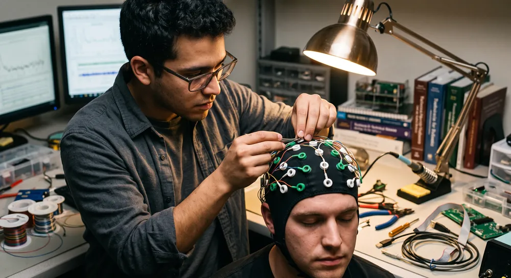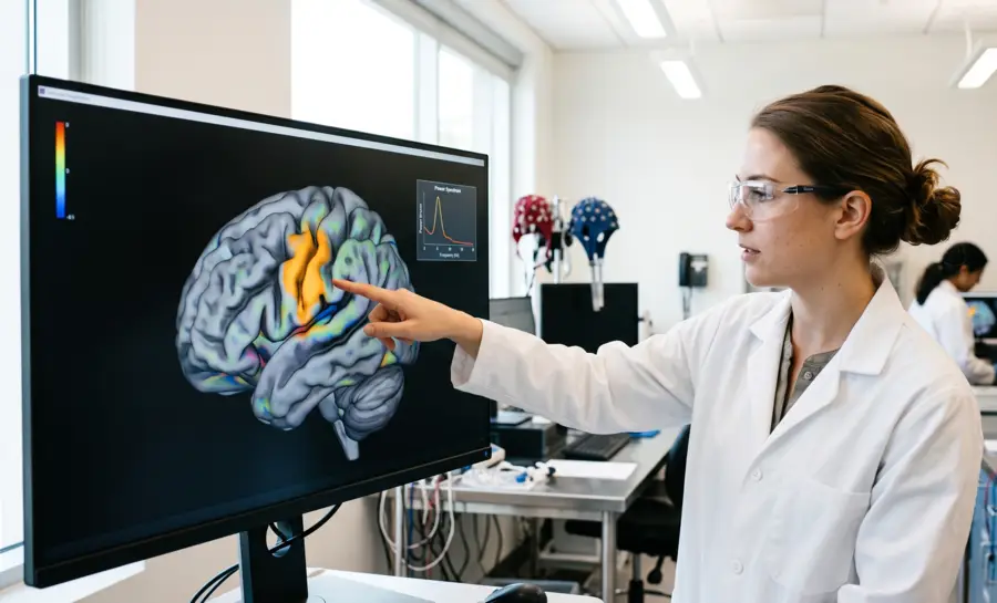
Among the different brain rhythms, one has captured the attention of neuroscientists for decades because it appears to sit at the intersection of action, perception, and social understanding.
The mu rhythm, an 8–13 Hz oscillation recorded over the sensorimotor cortex, decreases in power whenever we perform an action, watch someone else perform that same action, or even just imagine performing it. This property, known as desynchronization, has made the mu rhythm a central player in research on imitation, empathy, and clinical disorders ranging from stuttering to autism.
What Is the EEG Mu Rhythm?
The EEG mu rhythm is a patterned oscillation recorded from scalp electrodes over the sensorimotor regions of the brain. It is often described as an arch-shaped or comb-like rhythm with an alpha-like frequency, most commonly around 8–13 Hz, although reported boundaries vary across studies. The rhythm may be present during quiet wakefulness and can change when the sensorimotor system is engaged.
Mu activity is not a separate electrical substance or a direct readout of a single mental process. It is a measurable pattern in an electroencephalogram, shaped by neural sources, electrode placement, reference choice, and the person's state. A visible rhythm therefore has to be interpreted in context rather than treated as a standalone sign of a particular behavior or condition.
Mu Rhythm EEG: Where It Comes From
The mu rhythm originates in premotor and motor regions of the cortex and is continuously shaped by subcortical structures, particularly the basal ganglia, a collection of nuclei deep in the brain that regulate movement initiation and inhibition. The rhythm's sensitivity to both motor and sensory processes stems from its division into two functionally distinct frequency bands.
The slower component, mu-alpha, occupies the classic 8–13 Hz range and responds primarily to inhibitory signals from the basal ganglia and to sensory feedback arriving at the motor cortex. Think of mu-alpha as a gauge of how much the brain's motor output is being held in check and how incoming sensory information—the feel of a surface, the position of a limb—is being integrated.
The faster component, mu-beta, spans 14–25 Hz and reflects a different computational stream. It captures the brain's internal forward models, the predictions the motor system makes about the sensory consequences of movement. When you reach for a cup, your brain anticipates the tactile sensation before your fingers make contact. Mu-beta tracks the timing and precision of these motor-to-sensory projections.
This dual-band architecture means that researchers can observe simultaneous changes in mu-alpha and mu-beta across the time course of specific events, creating a rich window into neurophysiological function. Disruptions in one band but not the other can reveal whether a clinical condition stems from faulty inhibition, degraded sensory feedback, or disrupted internal timing.
For instance, in people who stutter, mu-alpha suppression during a working memory task is significantly reduced compared to fluent speakers, pointing toward specific basal ganglia and sensorimotor integration deficits that unfold with high temporal precision.
Feature | Mu-Alpha | Mu-Beta |
|---|---|---|
Frequency | 8–13 Hz | 14–25 Hz |
Main role | Inhibition and sensory feedback | Internal forward model predictions |
Sensitive to | Basal ganglia signals | Motor-to-sensory projection timing |
Clinical example | Reduced suppression in stuttering | Disrupted internal timing |
How to Identify the Mu Rhythm
On a raw EEG tracing, mu rhythm may appear as a relatively regular, arching waveform over central leads. It can be bilateral or stronger on one side, and it may be intermittent rather than continuously visible. Its shape can appear sharply contoured without being epileptiform, so morphology alone should not determine clinical meaning.
A practical identification process compares the suspected rhythm with its scalp distribution, frequency, state dependence, and response to a relevant task. A rhythm that changes with contralateral hand movement or another sensorimotor manipulation is more consistent with reactive mu activity than an equally shaped rhythm that remains unchanged. Even then, the finding is probabilistic rather than conclusive.
Several observations are particularly useful when reviewing a recording:
The activity is maximal or prominent near central electrodes rather than exclusively posterior electrodes.
Its dominant frequency falls within an alpha-like sensorimotor range.
Its amplitude or power changes during movement, motor imagery, or action observation.
The pattern is reproducible across trials and is not explained by muscle or electrode artifact.
These checks reduce the risk of confusing mu with posterior alpha, rhythmic artifact, or an incidental normal variant. They also show why a single short segment of EEG rarely answers the full interpretive question.
Mu Rhythm vs Alpha Rhythm: Key Differences
Mu and alpha rhythms can overlap in frequency, which is the main reason they are sometimes confused. The distinction is not made by frequency alone. Alpha is typically associated with posterior visual regions and changes with eye opening or closing, whereas mu is associated more closely with central sensorimotor regions and changes with sensorimotor engagement.
Their waveforms can also look similar. Both may be rhythmic, and both can vary in amplitude across states and people. A central rhythm that resembles alpha should be tested for topography and reactivity rather than classified solely from its appearance.
The following contrasts are useful but not absolute:
Question | Mu rhythm | Alpha rhythm |
|---|---|---|
Where is it usually strongest? | Central sensorimotor scalp regions | Posterior or occipital scalp regions |
What commonly changes it? | Movement, motor imagery, or action observation | Visual state, especially eyes open versus closed |
What does frequency tell us? | It may occupy an alpha-like range | Frequency alone is not sufficient for identification |
What is the main interpretive risk? | Confusing it with posterior alpha or artifact | Confusing posterior activity with central mu |
These distinctions guide analysis but do not replace examination of the full montage. Reference selection, volume conduction, and overlapping sources can make scalp distributions less clean than textbook examples suggest.
EEG Mu Rhythm Analysis Methods
EEG mu studies use both visual and quantitative methods. Visual review considers waveform shape, distribution, state, and reactivity, while quantitative analysis estimates power or amplitude in a preselected frequency range. Reliable work usually reports the reference, filters, artifact exclusions, epoch timing, baseline, and statistical model so that the result can be evaluated and reproduced.
Time-frequency analysis is often useful because mu responses may be transient and task-locked. Frequency analysis converts voltage changes into a representation of rhythmic components, while time-resolved methods show when a change begins and how long it lasts. Researchers must still distinguish a genuine neural effect from leakage across frequencies, filtering effects, and non-neural contamination.
Event-Related Desynchronization Explained
Event-related desynchronization, or ERD, is a task-related reduction in oscillatory power relative to a reference period. For mu studies, ERD is commonly calculated around movement or motor imagery and expressed as a percentage or decibel change from baseline. The method captures a relative response, which is often preferable to comparing raw amplitudes across people with different skull conductivity, electrode contact, or baseline power.
A typical ERD analysis divides the recording into trials, removes or marks contaminated segments, estimates spectral power, and compares a task interval with a prestimulus or resting interval. The exact result depends on window length, frequency resolution, baseline selection, and correction for multiple comparisons. Short windows preserve timing but give less stable frequency estimates; longer windows improve frequency precision but can blur rapid changes.
Interpretation should therefore include the full analysis design. A statistically significant reduction is evidence of a task-related signal change under those conditions, not proof that a particular psychological mechanism caused it. Replication across tasks, sessions, and samples strengthens the inference.
The Mirror Neuron Hypothesis and Its Discontents
The mu rhythm rose to prominence in social neuroscience because of an elegant hypothesis: if the same population of neurons fires when we act and when we observe action, and if the mu rhythm reflects that neural activity, then mu suppression during action observation could serve as a non-invasive index of mirror neuron system function. This logic drove hundreds of studies linking mu desynchronization to imitation, empathy, language development, and theory of mind.
The hypothesis rests on a firm empirical foundation. The mu rhythm does attenuate during both action execution and action observation, a property that distinguishes it from the posterior alpha rhythm, which responds primarily to visual attention. This functional similarity to mirror neurons, first discovered in the premotor and parietal cortices of macaque monkeys, made the mu rhythm an attractive tool for translational human research.
However, the specificity of this link has come under scrutiny. A critical test used crossmodal pattern classification to determine whether mu rhythm activity during action observation actually carries motor information or whether it instead reflects somatosensory features.
Participants observed actions that varied in terms of grip type (whole-hand or precision grip), the presence or absence of tactile stimulation, and whether the action involved using an object (transitive) or not (intransitive). When classifiers were trained to distinguish these action features, above-chance classification emerged for tactile stimulation and action transitivity, but tellingly, not for the motor detail of grip type.
The mu rhythm appeared to encode whether an object was being touched and whether a tool was involved, rather than the specific motor program required to execute the grasp.
This finding reframes the mu rhythm's functional role. During action observation, what desynchronizes over sensorimotor cortex may be a somatosensory representation, the brain's simulation of what the action would feel like, rather than a direct motor simulation of how to perform it.
The distinction matters because it constrains what kinds of inferences researchers can draw when they use mu suppression as a dependent measure. If the signal primarily carries tactile and transitivity information, then interpreting mu suppression during a social cognition task as evidence of motor mirroring becomes problematic without additional converging evidence.
Motor Experience Tunes the Sensorimotor Response
Despite these concerns about specificity, one finding consistently emerges: the mu rhythm is exquisitely sensitive to an individual's motor history. The brain's sensorimotor circuits do not respond uniformly to all observed actions. They resonate more strongly with actions that the observer has personally performed.
A direct test of this principle trained two groups of adults on a relatively novel action, using a claw-like tool to pick up a toy. One group received active motor training, physically manipulating the tool themselves until they could execute the action fluently. The second group received equivalent visual exposure, repeatedly watching and coding videos of the same action, but never performing it. A third group of complete novices had no prior experience with the tool at all.
Active motor training: physically manipulated the tool until fluent execution.
Visual exposure: watched and coded videos of the action, never performed it.
Complete novices: no prior experience with the tool at all.
When EEG was later recorded while all three groups watched the tool-use action, the performers showed the greatest mu rhythm desynchronization, particularly over the right hemisphere.
The observers, despite their extensive visual familiarity, showed a weaker response. The novices showed the weakest response of all.
This gradation demonstrates that active motor experience, more than passive observation, sculpts the sensorimotor circuits that generate the mu rhythm. The finding has significant implications for understanding imitation and early social learning. It suggests that when infants or adults acquire new motor skills, their brains become selectively tuned to detect and process those same actions when performed by others, a neural mechanism that could support the gradual building of a motor vocabulary that underlies imitation.
Notably, this study involved relatively short training periods, on the order of a single experimental session, rather than the years of expertise examined in classic mirror neuron research with dancers or athletes. The mu rhythm's sensitivity to brief motor learning episodes makes it a practical tool for studying how experience-dependent plasticity unfolds in the sensorimotor system.
From Motor Resonance to Emotional Insight
If mu suppression were merely a low-level motor resonance signal, its relevance to complex social cognition would be limited. But evidence now suggests that sensorimotor simulation, as measured by mu desynchronization, contributes to higher-order inferences about what other people are feeling.
In a pair of experiments that combined naturalistic video stimuli with continuous emotion ratings, participants watched clips of people describing emotional life events. Some participants saw the clips with both video and sound, some with only video, and some with only audio.
They provided moment-by-moment ratings of how they believed the target person felt, and empathic accuracy was operationalized as the correlation between the participant's ratings and the target's own self-reported emotional experience.
The critical finding was that right-lateralized mu suppression over sensorimotor regions tracked empathic accuracy. When participants showed stronger mu desynchronization, their emotional inferences more closely matched the target's actual feelings.
This relationship replicated across two cultural samples, one in the United States and one in Israel, though in the Israeli sample it held specifically when individualized frequency bands were used and only for the visual stimuli.
The modality-specific effect is theoretically important. It indicates that sensorimotor simulation contributes to empathic accuracy precisely when visual cues, facial expressions, gestures, and postural information, are available to drive the simulation. When only auditory information is present, other neural systems appear to carry the computational burden.
These results extend the functional significance of mu suppression well beyond low-level motor resonance. The sensorimotor cortex appears to participate in constructing the rich, contextualized inferences about others' internal states that constitute everyday empathy. The right-hemisphere lateralization aligns with broader findings in social neuroscience linking the right hemisphere to emotion processing and self-other discrimination.
Motor Imagery and the Search for Reliable BCI Control
The mu rhythm's responsiveness to imagined movement has made it a cornerstone signal for motor imagery brain-computer interfaces (BCIs). These systems translate the power changes that occur when a user imagines moving their left versus right hand into control commands for external devices. The principle states that an imagined hand movement suppresses the mu rhythm over the contralateral sensorimotor cortex, and this lateralized suppression can be detected and classified in real time.
BCI performance, however, varies substantially across individuals. In a 2024 study, authors mentioned that a significant minority of users, perhaps 15 to 30 percent, cannot achieve reliable control, a phenomenon termed BCI inefficiency. Identifying factors that predict performance has been a persistent goal of the field. Gender has been proposed as one such factor, with some earlier studies suggesting that females might show greater mu modulation during motor imagery tasks.
The large-scale 2024 investigation combined four independent datasets to assemble a sample of 248 participants with equal gender distribution, substantially larger than any prior study on the question. All participants performed a standard left-hand versus right-hand motor imagery task.
The analysis extracted the Mu Suppression Index from electrodes positioned over the left and right sensorimotor cortices and compared values between female and male participants. The result showed no significant difference emerged between groups. Gender, at least in this well-powered sample, did not predict the ability to modulate the mu rhythm during motor imagery.
This null finding has practical implications. BCI researchers and clinicians can likely set aside gender as a screening variable when recruiting participants or selecting users for motor imagery-based systems. The search for reliable predictors of BCI performance might need to focus elsewhere, perhaps on neurophysiological traits, cognitive strategies, or training protocols.
Clinical Signatures in Stuttering
The dual-band architecture of the mu rhythm makes it particularly suited to capturing the specific neural deficits that characterize certain clinical disorders.
Stuttering provides a compelling case study. The condition has long been associated with dysfunction in basal ganglia circuits that regulate the timing and initiation of movement, along with disrupted sensorimotor integration in speech production. Mu-alpha, sensitive to basal ganglia inhibitory signals and sensory-to-motor feedback, and mu-beta, sensitive to timing and forward model projections, offer complementary windows into these deficits.
In speech production tasks, independent component analysis can extract mu rhythms from raw EEG signals and map changes in alpha and beta power to the myogenic activity of the articulators. Studies using this approach have demonstrated that mu-alpha and mu-beta activity reliably differentiates people who stutter from fluent speakers during both speech and auditory discrimination tasks.
The temporal precision of EEG allows researchers to observe when, during the planning and execution of an utterance, the sensorimotor deficits emerge. A novel finding from a non-word repetition task further showed reduced mu-alpha suppression in a stuttering group compared to a typically fluent group, extending the mu rhythm's sensitivity to cognitive domains beyond speech itself.
What the Mu Rhythm Reveals About Action, Empathy, and Brain Signals
The mu rhythm's story is one of refinement, showing that a brain wave once labeled a mirror neuron index actually carries richer, more sensory information. When we watch someone act, the desynchronization we measure reflects what the action would feel like, not just the motor commands, a distinction that changes how researchers interpret social neuroscience data.
Personal motor experience sharpens this response, meaning the brain tunes itself to recognize actions it has physically practiced, which supports the gradual building of imitation skills. Meanwhile, the same signal predicts how accurately people read emotions from facial expressions across different cultures, extending the mu rhythm's relevance well beyond low-level movement.
The mu rhythm holds its value not through any single fixed interpretation but through a growing body of evidence that continues to sharpen what it measures and why that matters for human physiology.
References
Coll, M. P., Press, C., Hobson, H., Catmur, C., & Bird, G. (2017). Crossmodal Classification of Mu Rhythm Activity during Action Observation and Execution Suggests Specificity to Somatosensory Features of Actions. The Journal of neuroscience : the official journal of the Society for Neuroscience, 37(24), 5936–5947. https://doi.org/10.1523/JNEUROSCI.3393-16.2017
Cannon, E. N., Yoo, K. H., Vanderwert, R. E., Ferrari, P. F., Woodward, A. L., & Fox, N. A. (2014). Action experience, more than observation, influences mu rhythm desynchronization. Plos one, 9(3), e92002. https://doi.org/10.1371/journal.pone.0092002
Genzer, S., Ong, D. C., Zaki, J., & Perry, A. (2022). Mu rhythm suppression over sensorimotor regions is associated with greater empathic accuracy. Social Cognitive and Affective Neuroscience, 17(9), 788-801. https://doi.org/10.1093/scan/nsac011
von Groll, V. G., Leeuwis, N., Rimbert, S., Roc, A., Pillette, L., Lotte, F., & Alimardani, M. (2024). Large scale investigation of the effect of gender on mu rhythm suppression in motor imagery brain-computer interfaces. Brain computer interfaces (Abingdon, England), 11(3), 87–97. https://doi.org/10.1080/2326263X.2024.2345449
Jenson, D., Bowers, A. L., Hudock, D., & Saltuklaroglu, T. (2020). The application of EEG mu rhythm measures to neurophysiological research in stuttering. Frontiers in human neuroscience, 13, 458. https://doi.org/10.3389/fnhum.2019.00458
Frequently Asked Questions
What is the mu rhythm and why is it important in neuroscience?
The mu rhythm is a rhythmic electrical oscillation recorded over the sensorimotor cortex that decreases in power whenever a person performs, observes, or imagines an action. This property, called desynchronization, makes it a key signal for studying action, perception, and social understanding.
How is the mu rhythm divided into functional components, and what does each one do?
The mu rhythm is divided into a slower component and a faster component. The slower component responds to inhibitory signals from the basal ganglia and sensory feedback, while the faster component tracks the brain's internal predictions about the sensory consequences of movement.
Does the mu rhythm have any connection to empathy or understanding other people's feelings?
Yes, stronger mu suppression over the right sensorimotor region tracked with higher empathic accuracy when participants watched emotional videos. This relationship held specifically when visual cues like facial expressions and gestures were available, indicating the mu rhythm supports inferences about others' internal states.
Can the mu rhythm be used in brain-computer interfaces?
The mu rhythm is a cornerstone signal for motor imagery brain-computer interfaces because imagined movement causes lateralized suppression that can be translated into commands.
Emotiv is a neurotechnology leader helping advance neuroscience research through accessible EEG and brain data tools.
Medical Disclaimer: The information provided on this website is for educational and informational purposes only and is not intended as medical or health advice. This content may contain errors and should not be relied upon to make life-altering health, medical, or lifestyle choices. Always seek the advice of your physician or other qualified health provider with any questions you may have regarding a medical condition or treatment.
Christian Burgos