When you look at an EEG readout, you are looking at a set of choices, not just raw data pulled from the scalp. Before a single waveform appears on screen, a technician or software system has already decided which electrodes get compared to which. That decision framework is called a montage, and it shapes everything a clinician or researcher sees.
Understanding this concept is a necessary step before diving into any specific electroencephalogram (EEG) reading, because the same set of electrodes can produce dramatically different-looking traces depending on how they are paired.
What is an EEG Montage?
An EEG recording involves capturing electrical potentials from the scalp to visualize brain activity. To make sense of this information, practitioners use specific display configurations called montages, which act as the lenses through which they view neural signals.
These arrangements are essential for standardized interpretative processes across clinical and research environments.
Why Raw Voltages Need a Comparison Point
An electrode on the scalp records a voltage, but that number is not meaningful on its own. Voltage is inherently relative. Without a second point of comparison, there is no way to know if a given reading reflects genuine brain activity or simply electrical drift, movement, or noise from the recording equipment itself.
This is why every EEG channel is built as a differential measurement. The waveform displayed for any given channel is the electrical activity recorded at one electrode minus the activity recorded at a second electrode.
This subtraction step is the reason EEG works at all in a noisy clinical or research environment. If two nearby electrodes both pick up the same distant interference, say from a piece of equipment in the room, that shared interference cancels out when one signal is subtracted from the other.
Engineers refer to this as common-mode rejection, a plain way of saying that anything common to both electrodes gets filtered out automatically, leaving behind only the differences that are more likely to reflect local brain electrical activity. Every montage, no matter how it is designed, depends on this differential principle.
The choice of which two points to compare is what changes between montage types, but the underlying math of subtraction stays constant.
EEG channels are differential measurements: one electrode's voltage subtracted from another.
Raw voltages lack meaning without a reference point; comparisons distinguish brain activity from noise.
Common-mode rejection cancels shared interference, a principle that makes EEG viable in noisy settings.
Every montage relies on this subtraction; only the choice of electrode pairs varies.
How EEG Electrode Placement Works
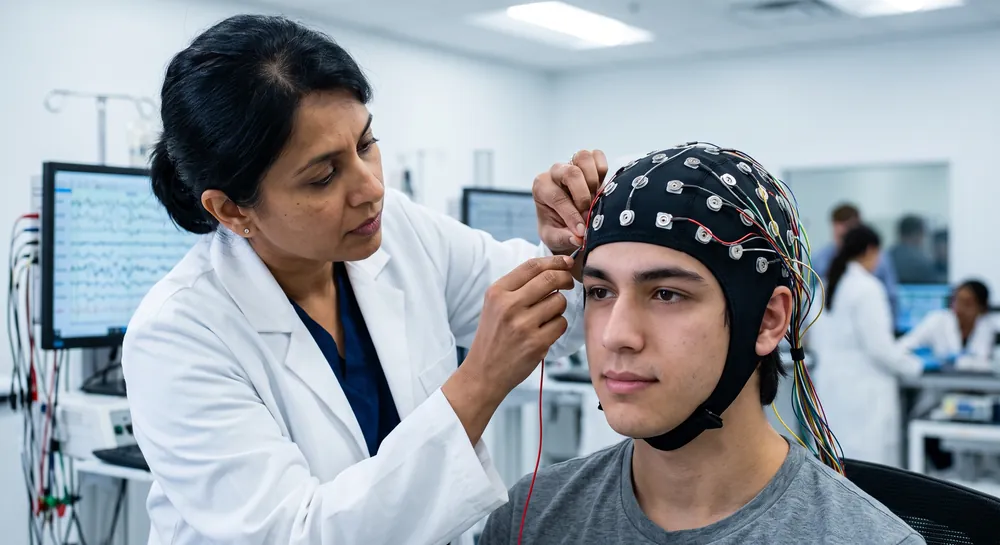
Technicians typically follow standardized procedures to ensure reproducibility when connecting scalp sensors to the amplifier. This measurement process relies on the relationship between physical landmarks such as the nasion and the inion to verify that each sensor sits in an anatomically accurate location.
Such consistent physical landmark protocols allow clinicians and researchers to compare results across different sessions or even across different facilities.
Why are EEG Montages Important?
Electrode configurations are vital for transforming raw voltage inputs into readable diagnostic data. By grouping electrodes in specific spatial clusters, the chosen arrangement can highlight localized discharges that might otherwise remain obscured by global activity.
Detecting Seizures and Abnormal Activity
When investigating potential epileptiform activity, selecting a configuration that maximizes spatial contrast is critical for identifying local neuronal irritability.
A sensitive setup often reveals sharp waves or spikes which are the hallmarks of localized brain dysfunction. During neuroscience evaluations, this spatial resolution helps correlate observed behaviors with specific scalp distribution patterns.
Diagnosing Neurological Disorders
Clinicians rely on various recording patterns to establish a differential diagnosis for conditions ranging from degenerative disorders to metabolic encephalopathies. The ability to see both focal and generalized signals enables a comprehensive assessment of the patient's state throughout the study.
This rigorous clinical standard documentation provides the foundation for determining the underlying cause of neurological shifts in patients.
Measuring Brain Function
Continuous monitoring allows for the observation of evolving patterns over time, providing insight into the stability of neural output in patients under sedation or physiological stress. By monitoring specific cortical regions, staff can identify subtle changes in the depth of processing or the emergence of rhythmic activities that indicate distress.
Types of EEG Montages Explained
There are several ways to categorize the display of brain potentials to optimize the diagnostic yield of any given recording session. Practitioners must select the approach that best suits the question at hand, whether it requires focusing on local features or characterizing broader background patterns.
Bipolar and Referential Approaches to Building Channels
Montages generally fall into two broad families.
A bipolar montage links adjacent electrodes together in a chain, so each channel reflects the voltage gradient between two neighboring points on the scalp. This approach tends to highlight sharp, localized differences in activity because it is only ever comparing electrodes that are physically close to one another.
A referential montage takes a different approach. Instead of comparing neighbors, every electrode is measured against one shared reference point, which might be a single electrode near the ear, or a mathematical average built from all the electrodes on the scalp.
This produces a broader picture of activity across the head, but it comes with a catch: the entire recording becomes dependent on how neutral that single reference point actually is. If the reference itself carries some hidden electrical activity, that activity gets subtracted into every single channel, distorting the picture of where brain activity is truly concentrated.
This is why finding a genuinely neutral reference point remains an active area of investigation rather than a solved problem.
Research comparing common re-referencing methods, including linked-mastoid referencing, average referencing, and a technique called the reference electrode standardization technique (REST), has found that both average referencing and REST produce comparatively low reconstruction errors relative to linked-mastoid referencing. REST in particular showed less sensitivity to artifacts mixed into the recording.
Critically, the accuracy of these methods depends heavily on two other factors:
How many electrodes are used
Whether the calculations rely on a realistic model of the head's shape rather than a simplified sphere
A high-density electrode montage combined with a realistic head model substantially improved the reliability of estimating a neutral reference point, which in turn improves the accuracy of every channel built from that reference.
Bipolar Montage | Referential Montage |
|---|---|
Compares adjacent electrodes in a chain | Measures against one shared reference |
Highlights sharp, localized voltage gradients | Shows broader scalp activity |
Good for localized differences | Depends on neutral reference point |
The Double Banana EEG Montage Pattern
This classic layout is a standard configuration that uses two parallel lines of electrodes stretching from anterior to posterior regions, creating pairs that resemble the curve of a banana on both sides of the head. It is widely favored because it covers all major cortical regions efficiently, making it the go-to standard for identification of hemispheric differences.
Laplacian Montage EEG Analysis
This method applies a mathematical transformation to the data to sharpen the signal and reduce the influence of volume conduction from distant sources.
It effectively enhances the representation of activity directly beneath the electrode cluster while diminishing noise from deeper or adjacent structures. This is a highly technical evaluation technique used primarily in advanced research and specific complex diagnostic cases.
The EEG 10 20 Montage System
Consistency is the cornerstone of clinical diagnostic reliability, particularly when multiple specialists are assessing images from the same patient. The standardized spacing system dictates the exact placement of electrodes, ensuring that the findings are always relatable to the same anatomical lobes.
Electrode Placement in the 10-20 System
Technicians follow a structured 10-20 protocol to maintain uniformity across all studies, relying on the following key points:
The distance from the inion to the nasion serves as the midline anchor.
Electrodes are spaced by 10% or 20% intervals along the skull.
Odd-numbered sensors represent the left hemisphere locations.
Even-numbered sensors define the right hemisphere surface areas.
Using this system prevents ambiguity in localization, as it compensates for variations in head circumference and shape. This standardized anatomical mapping system ensures that the results remain reliable regardless of the technician performing the setup or the equipment used for recording.
Choosing the Right EEG Montage for Your Needs
Selecting the optimal recording setup requires a clear understanding of the clinical question or research goal. If the objective is to localize a focal seizure, bipolar arrangements generally provide the highest diagnostic yield due to the localized spatial filtering effect. Researchers often start with a screening configuration like the double banana before cycling through other modes to narrow down specific regions of interest.
When the primary interest involves global brain state changes, such as in metabolic disturbances, a referential arrangement typically offers a more accurate representation of the signal distribution. This allows practitioners to observe voltage variations across the entire scalp without the phase reversals seen in bipolar traces. Relying on a predefined protocol selection method consistently yields superior results in professional clinical settings.
Effective interpretation stems from the strategic use of multiple modalities during a single session, rather than relying on one solitary viewing format. By comparing data across various configurations, practitioners can gain confidence in their localizing assessments and ensure no subtle abnormality is missed. A methodical approach ensures the best information is available for clinical decision-making processes.
Tailoring Montages to Specific Research and Clinical Questions
Montages are not fixed templates handed down without adjustment. They can be simplified, expanded, or optimized depending on what a specific study or clinical situation demands.
In research combining EEG with functional near-infrared spectroscopy (fNIRS), a technique that measures blood flow changes linked to brain activity, investigators developed a method for computing an optimal arrangement of light sources and detectors on a cap.
Rather than using a fixed, whole-head sensor layout, this method mathematically identifies the placement that maximizes sensitivity over a specific brain region relevant to an individual patient's epileptic activity. When tested, this customized montage achieved spatial resolution comparable to standard whole-head arrangements while using significantly fewer sensors, along with an improved signal-to-noise ratio over the targeted region.
This illustrates a core principle behind montage customization: fewer sensors do not automatically mean less useful data, provided their placement and pairing are calculated with a specific question in mind.
Moreover, speed and simplicity matter just as much in intensive care settings, where a full 21-channel EEG setup can be impractical for a critically ill patient who needs rapid assessment.
One 2022 study tested a 10-electrode montage applied at the bedside by an intensivist who had completed a year-long electrophysiology training course. Compared against an expert neurophysiologist's readings, this trained intensivist reached acceptable agreement on several key patterns:
94 percent for minimum background frequency
89 percent for maximum background frequency
100 percent for burst suppression
83 percent for background continuity
When 22 non-expert intensivists went through only a single one-hour training session, their reliability was more mixed, though most still reached acceptable agreement for background frequency measures. This points toward a workable middle ground for critical care, where a reduced montage paired with focused training can produce clinically useful readings without the full setup time of a conventional system.
How Electrode Density Shapes Signal Quality
More electrodes generally provide more spatial detail and more accurate referencing, but that comes at the cost of setup time, patient tolerance, and expense. The relevant research maps this trade-off with some precision.
The study comparing re-referencing methods found that high electrode density reduced reconstruction errors for both average referencing and REST. Interestingly, the two methods performed differently depending on density.
With a low-density montage, REST produced a more reliable estimate of a neutral reference than average referencing did. With a high-density montage, average referencing performed just as well, unless precise information about electrode positions on the head was unavailable, in which case REST retained an advantage.
Further, the 2022 postanoxic coma study aforementioned reached a related conclusion from the clinical side, noting that limited-channel EEG can serve as a cost-effective alternative to full 21-channel monitoring. That said, its data revealed a specific weak point: maximum background frequency, a subtler measure of brain activity patterns, was correctly classified in only 70 percent of cases when using the reduced montage.
This suggests that while a limited montage captures the major clinical patterns, finer-grained frequency details are more likely to be lost as electrode count drops.
Where Montage Choices Create Risk of Misinterpretation
Every montage design carries trade-offs, and the research reviewed here points to specific, well-documented pitfalls rather than a vague warning against simplification. The clearest example comes from the postanoxic coma study's 31 percent false positive rate for periodic discharges, traced directly to eye movement artifacts recorded over the bifrontal electrodes.
This happened because a limited-channel setup lost some of the spatial context that a fuller montage provides, context that would normally help a reader distinguish an eye movement from genuine periodic brain activity. Fewer channels mean fewer independent viewpoints on the same signal, and that reduction in perspective is what allows certain artifacts to masquerade as pathology.
Reference choice introduces a comparable risk on the referential side of montage design. The re-referencing study demonstrated that an inappropriate reference point, such as linked mastoids under certain conditions, can distort the apparent distribution of activity across the scalp.
It also showed that relying on a simplified spherical head model rather than a realistic one worsens the performance of REST specifically, since the mathematics behind REST depends on an accurate representation of how electrical activity actually travels through the layers of the head.
These findings function as known, well-documented challenges that inform careful practice. None of them amount to a case against simplified EEG altogether. They simply mark where extra caution and cross-checking are warranted.
Montages as a Flexible, Logical Toolkit
An EEG montage is, at its core, a set of rules for deciding which electrode pairs become channels. That decision transforms a grid of individual sensors into waveforms that a clinician or researcher can actually interpret, whether the goal is spotting a seizure at the bedside or mapping activity across the neuroscience research setting of a lab.
There is no universal best montage, only a best montage for a given question. Bipolar chains are suited to picking out sharp, localized voltage gradients between neighboring points. Referential schemes, when paired with a carefully chosen and validated reference, support broader mapping of activity distributed across the scalp.
Customized or reduced montages, whether built for combined EEG-fNIRS work or for rapid ICU assessment, become valuable when speed, patient comfort, or cost outweigh the benefits of a full sensor array, provided the reduced layout is designed with the same rigor as a full one.
The studies reviewed here point toward a field still actively refining these trade-offs. High-density montages paired with realistic head models improve reference estimation, but low-density setups with the right reference method can still perform reliably in specific settings. Reduced electrode counts can preserve clinically meaningful information, but only when artifact risk and rater consistency are accounted for.
These remain open questions rather than settled conclusions.
Mastery of montage strategies acts as the foundation for high-quality neurophysiological analysis and clinical diagnosis. By understanding how to organize and view complex brain data, practitioners can ensure consistent and actionable interpretations that support clear diagnostic pathways for patients.
References
Liu, Q., Balsters, J. H., Baechinger, M., Van der Groen, O., Wenderoth, N., & Mantini, D. (2015). Estimating a neutral reference for electroencephalographic recordings: the importance of using a high-density montage and a realistic head model. Journal of neural engineering, 12(5), 056012.
Abid, S., Papin, G., Vellieux, G., de Montmollin, E., Wicky, P. H., Patrier, J., ... & Sonneville, R. (2022). A simplified electroencephalography montage and interpretation for evaluation of comatose patients in the ICU. Critical Care Explorations, 4(11), e0781. https://doi.org/10.1097/CCE.0000000000000781
Frequently Asked Questions
What exactly is an EEG montage, and how does it differ from an electrode placement map?
A montage is the rulebook that defines which electrodes are paired together to create each channel, showing the voltage difference between two recording sites. An electrode placement map like the 10-20 system only describes sensor locations on the head, while the montage determines how those signals are combined to produce readable waveforms.
Why can’t we just read the voltage from a single electrode without comparing it to another?
Voltage is a relative measure, so a single reading has no meaningful context and can be influenced by drift or noise. EEG uses differential measurement, subtracting one electrode’s signal from another to cancel out common interference and reveal local brain activity.
What is the difference between a bipolar and a referential montage?
A bipolar montage links adjacent electrodes in a chain, comparing neighbors to highlight sharp, localized voltage differences. A referential montage compares every electrode to one shared reference point, giving a broader view of scalp-wide activity but making the recording dependent on the neutrality of that reference.
How does the choice of reference electrode affect an EEG reading?
If the reference site carries its own electrical activity, that activity gets subtracted into all channels, distorting the scalp distribution of brain signals. Methods like average referencing or REST attempt to estimate a neutral reference, but accuracy depends on electrode density and head model realism.
Can a reduced number of electrodes still provide clinically useful information?
Yes, carefully designed reduced montages can preserve key patterns like background continuity or seizure detection, especially when tailored to a specific question or setting. However, losing channels can reduce spatial context, making it harder to distinguish artifacts from genuine activity.
What are the main risks of misinterpretation when using a limited montage?
A common risk is that eye movement artifacts can be mistaken for abnormal brain activity because fewer channels offer less independent viewpoints. Additionally, an inappropriate reference or differences in how raters interpret the data can further complicate readings.
Does more electrodes always mean better data quality?
Higher density generally improves spatial detail and reference accuracy, but it’s not the only factor; the arrangement of electrodes and consistency of interpretation matter just as much. In some focused applications, a well-designed reduced montage can perform comparably to a full setup.
Is there one best montage for all EEG recordings?
No universal best montage exists; the optimal choice depends on the clinical or research question. Bipolar chains suit detecting local voltage gradients, referential schemes enable broad mapping, and customized montages balance speed and sensitivity for specific tasks.
Can artificial intelligence affect montage interpretation?
While software automates the display process, human expertise remains necessary to verify the clinical context and distinguish between genuine pathological activity and technical artifacts.
Emotiv is a neurotechnology leader helping advance neuroscience research through accessible EEG and brain data tools.
Medical Disclaimer: The information provided on this website is for educational and informational purposes only and is not intended as medical or health advice. This content may contain errors and should not be relied upon to make life-altering health, medical, or lifestyle choices. Always seek the advice of your physician or other qualified health provider with any questions you may have regarding a medical condition or treatment.
Christian Burgos