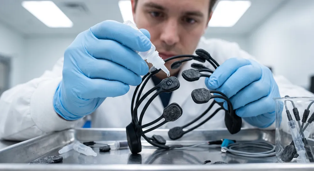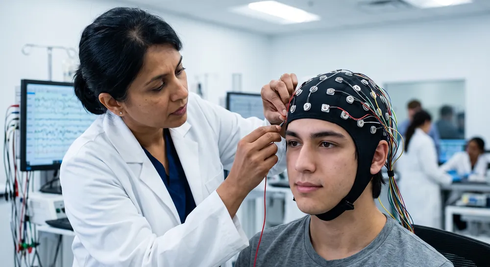
Defined as rhythmic neural activity falling between roughly 13 and 30 Hz, beta waves stand apart from slower waves associated with drowsiness and deep rest. Where delta and theta rhythms mark a brain winding down, beta activity is associated with a brain switching on. Electroencephalogram (EEG) recordings capture these patterns through electrodes on the scalp, revealing a frequency band that appears when a person focuses, exerts cognitive effort, or prepares to move.
What Are Beta Brain Waves?
Beta brain waves are patterns of rhythmic electrical activity associated with waking consciousness and active mental engagement. They are not a single, uniform signal; beta activity can vary in frequency, strength, location, and timing.
Researchers study these patterns within the broader field of neuroscience, where brain activity is related to cognition, movement, emotion, and health. A beta reading is therefore best understood as one feature of a larger neurophysiological picture.
The Frequency Range of Beta Brain Waves
Beta frequency is commonly described as approximately 13–30 hertz, meaning 13 to 30 cycles per second. Some references use slightly different boundaries, such as 12–30 Hz or 14–38 Hz, because frequency bands are conventions rather than sharply separated biological categories.
An EEG records voltage changes at the scalp, allowing researchers and clinicians to examine the distribution and timing of these rhythms.
Where Beta Waves Originate in the Brain
Beta oscillations appear most prominently in specific brain regions. Sensorimotor cortices, the basal ganglia, and the prefrontal cortex all generate beta activity, though the exact source remains unresolved.
One 2021 review notes that beta has been predominantly observed in sensorimotor cortices and basal ganglia structures, where it is thought to be involved in somatosensory processing and motor control. This suggests that the rhythm is a distinct feature of healthy sensorimotor function.
The question of where beta arises is not settled. A second 2019 review flags three possibilities: beta could be generated in the basal ganglia, in the cortex, or through a combined interaction between both regions.
Each option carries different implications for how beta supports cognition. A basal ganglia origin would tie beta closely to movement initiation and inhibitory circuits. A cortical origin would emphasize beta's role in local processing and long-range communication. A combined model would suggest beta emerges from coordination between subcortical timing structures and cortical processing layers.
This uncertainty does not diminish beta's importance. It does signal that beta likely performs different jobs in different circuits. The same frequency range may encode motor inhibition in one region while supporting memory reactivation in another. Acknowledging this regional heterogeneity helps explain why beta has resisted description by any single functional account.
How Beta Waves Support Attentional Selection
Experimental evidence directly links beta to the brain's selection of relevant information. In a visual oddball task, participants view a stream of stimuli and respond when a rare target appears.
One study measured EEG from 30 cortical locations while healthy older adults performed this task. The results showed that beta power, inter-trial phase synchronization, and event-related beta responses in the 15–20 Hz range were significantly higher for target stimuli than for non-target stimuli. This difference was especially pronounced at occipital electrode sites, which process incoming visual information.
These results suggest that increased beta in response to targets could shift the brain into an attention state, preparing cortical networks for deeper processing. Further, beta rose specifically when the stimulus carried behavioral relevance. This selectivity points toward beta as a possible operator in cognitive activity, not a passive reflection of visual stimulation.
In patients with mild cognitive impairment (MCI), the target versus non-target beta distinction disappeared. The evoked beta power in MCI patients showed no difference between relevant and irrelevant stimuli.
This loss of discrimination is consistent with the attentional difficulties that characterize early cognitive decline. It also suggests that a blunted beta response might serve to track the functional impact of early neurodegenerative processes. However, this is a single study with 17 participants per group, and the correlation requires replication before clinical application becomes feasible.
Beta Waves in Working Memory and Executive Control
Working memory tasks require the brain to hold information briefly, manipulate it, and discard it when no longer needed. Beta oscillations track each phase of this cycle.
According to one 2017 review, beta appears in short-lived, flexible bursts that support the endogenous reactivation of stored cortical representations. Rather than passively preserving a current state, these beta-mediated networks may actively wake a cognitive set to meet immediate task demands.
Moreover, the aforementioned 2019 review reports that PFC beta increases during the delay period of working memory tasks, when information must be held without external support. This delay-period beta might help maintain current contents or prevent interference from distraction. Both functions would support the same behavioral outcome: keeping the relevant item accessible while suppressing competing signals.
However, a more counterintuitive finding emerges at the end of a trial. When working memory information must be erased, PFC beta increases again.
The review calls this the clear-out function. The brain may use beta not only to hold information but also to actively remove it when the task ends. A similar pattern appears during the stopping of action and the stopping of long-term memory retrieval, where increased prefrontal beta also occurs. The clear-out hypothesis frames beta as a signal for deactivating no-longer-relevant content.
However, the review cautions that alternative interpretations exist. The clear-out in PFC might have a counterpart in the postmovement clear-out of motor plans in sensorimotor cortex, but recent studies support other explanations as well. Therefore, the precise mapping between PFC beta and sensorimotor beta remains an open question.
Beta Waves and Motor Control
One of beta's longest-standing associations is with movement. Sensorimotor cortices and basal ganglia structures are central to voluntary motor planning and execution, and beta activity is particularly strong in these regions.
During movement preparation, beta patterns reorganize. During and after movement, beta activity changes again. The rhythm appears tied to the sensorimotor loop that integrates touch, position awareness, and planned action.
Motor learning adds another dimension given that when people acquire new motor skills, beta activity shifts. Research suggests beta may help integrate sensory input with prior contextual knowledge, allowing the brain to update motor programs based on what it already knows.
This integration function could explain why beta appears during both the planning and the correction of movement. It may also help explain why beta is disrupted in conditions affecting motor control.
Beta Waves in Temporal Prediction
Predicting when an event will occur is central to adaptive behavior. A tennis player must anticipate the ball's arrival. A driver must time a lane change.
Beta oscillations appear to contribute to this temporal prediction system. Motor control and temporal prediction share a natural connection, they both require the brain to prepare an action before the action occurs.
One study by Arnal et al. tested this directly using a rhythmic listening task. Participants heard brief tone sequences and detected target delays while undergoing magnetoencephalography recording.
The results showed that delta oscillations (1–3 Hz) and beta oscillations (18–22 Hz) coupled together and temporally aligned with upcoming targets before the targets occurred. This coupling biased decisions toward correct responses. When delta and beta activity aligned well with the expected target time, participants were more likely to respond accurately.
The study interprets the delta-beta coupling as evidence that the motor system plays an active role in temporal prediction. The beta component, associated with motor preparation, appears to align with slower delta rhythms that track rhythmic structure. This coordination between sensory and motor systems could allow the brain to select relevant sensory information at precisely the right moment.
Beta's predictive activity happens prior to target onset, linking attentional preparation with motor timing. The brain is not passively waiting for events. It may be actively using oscillatory dynamics to anticipate them.
Too Much or Too Little Beta Activity: Potential Implications
Beta activity can be described as relatively high or low only in relation to a recording method, reference condition, and population. A change in beta power may reflect cognition, emotion, movement, muscle contamination, medication effects, sleep pressure, or technical factors.
For that reason, a numerical value alone does not establish a disorder. Clinical interpretation combines EEG findings with symptoms, history, examination, and other evidence.
Excessive Beta Waves and Anxiety
Periods of anxious arousal may involve persistent alertness, rapid thought, and difficulty relaxing, all of which can coincide with increased beta activity in some settings. The association is not universal, and elevated beta is not a diagnostic test for anxiety. High-frequency muscle activity can also be mistaken for cerebral beta, particularly around the forehead, jaw, and temples.
Researchers therefore distinguish genuine cortical signals from artifacts and examine whether a pattern is consistent across conditions. Anxiety assessment generally relies on clinical evaluation rather than on beta activity alone.
can contribute to research and selected diagnostic investigations, but it does not independently explain the cause of distress.
Low Beta Waves and Lack of Focus
Lower beta activity may occur during drowsiness, reduced engagement, or some states of diminished alertness. It can be associated with difficulty sustaining attention, but that relationship varies across individuals and experimental conditions. A low reading may also result from electrode placement, filtering choices, reference selection, or a recording taken during a naturally quiet state.
This is why frequency analysis is used to examine the full spectrum rather than one band in isolation. EEG frequency analysis can help organize delta, theta, alpha, beta, and gamma components, while interpretation still depends on signal quality and clinical context. Terms such as “low beta” describe an observation, not a diagnosis.
Why Understanding Beta Waves Matters for Brain Health
Across attention, memory, movement, and timing, beta waves appear when the brain is actively engaged. This pattern suggests the rhythm is less about maintaining a steady state and more about helping the brain filter distractions, hold and release information, prepare actions, and anticipate what comes next. The brain seems to use beta activity to ready itself for what matters.
These findings carry practical possibilities for brain health and neurotechnology. Altered beta responses to meaningful stimuli could offer an early warning sign of memory decline, and the timing of beta signals could help brain stimulation devices adjust in real time.
As neuroscience research shifts from treating beta as a continuous tone to reading its brief bursts, the picture of how this rhythm supports everyday thinking will likely grow sharper.
References
Barone, J., & Rossiter, H. E. (2021). Understanding the role of sensorimotor beta oscillations. Frontiers in systems neuroscience, 15, 655886. https://doi.org/10.3389/fnsys.2021.655886
Schmidt, R., Herrojo Ruiz, M., Kilavik, B. E., Lundqvist, M., Starr, P. A., & Aron, A. R. (2019). Beta oscillations in working memory, executive control of movement and thought, and sensorimotor function. The Journal of Neuroscience, 39(42), 8231-8238. https://doi.org/10.1523/JNEUROSCI.1163-19.2019
Güntekin, B., Emek-Savaş, D. D., Kurt, P., Yener, G. G., & Başar, E. (2013). Beta oscillatory responses in healthy subjects and subjects with mild cognitive impairment. NeuroImage: Clinical, 3, 39-46. https://doi.org/10.1016/j.nicl.2013.07.003
Spitzer, B., & Haegens, S. (2017). Beyond the status quo: a role for beta oscillations in endogenous content (re) activation. eneuro, 4(4), ENEURO-0170. https://doi.org/10.1523/ENEURO.0170-17.2017
Arnal, L. H., Doelling, K. B., & Poeppel, D. (2015). Delta–beta coupled oscillations underlie temporal prediction accuracy. Cerebral Cortex, 25(9), 3077-3085. https://doi.org/10.1093/cercor/bhu103
Frequently Asked Questions
What are beta oscillations and how are they defined in the brain?
Beta oscillations are rhythmic neural activity occurring between 13 and 30 Hz, detected via EEG electrodes on the scalp. They are associated with states of active cognitive engagement, such as focus, working memory, and motor preparation, rather than with drowsiness or deep rest.
Where in the brain are beta oscillations primarily generated?
Beta activity is most prominent in the sensorimotor cortices, basal ganglia, and prefrontal cortex. However, the exact generation site remains unresolved, with evidence supporting cortical origins, basal ganglia origins, or a combined interaction between both regions.
How do beta oscillations contribute to attentional selection?
Beta power increases specifically when the brain processes relevant stimuli, such as rare targets in a visual oddball task, especially at occipital sites. This selective enhancement suggests beta prepares cortical networks for deeper processing of behaviorally important information.
What role do beta oscillations play in working memory?
Beta appears in short bursts during the delay period of working memory tasks, helping to maintain relevant information and prevent distraction. At the end of a task, beta activity increases again to "clear out" no-longer-needed content, a process called the clear-out function.
How are beta oscillations linked to motor control and learning?
Beta activity is strong in motor regions during movement preparation and changes during and after movement, supporting planning and execution. It also shifts when learning new motor skills, helping the brain integrate sensory input with prior knowledge to update motor programs.
What is the significance of delta-beta coupling in temporal prediction?
Delta and beta oscillations align with expected event timing, biasing decisions toward correct responses. This coupling reflects the motor system's active role in predicting when events will occur, allowing the brain to select relevant sensory information at the right moment.
What are the potential clinical applications of beta oscillation research?
Beta signals could guide adaptive deep-brain stimulation by indicating when a cognitive episode ends, enabling real-time parameter adjustments. Additionally, blunted beta responses to targets may serve as a biomarker for early cognitive decline, aiding in earlier detection of conditions like mild cognitive impairment.
Emotiv is a neurotechnology leader helping advance neuroscience research through accessible EEG and brain data tools.
Medical Disclaimer: The information provided on this website is for educational and informational purposes only and is not intended as medical or health advice. This content may contain errors and should not be relied upon to make life-altering health, medical, or lifestyle choices. Always seek the advice of your physician or other qualified health provider with any questions you may have regarding a medical condition or treatment.
Christian Burgos