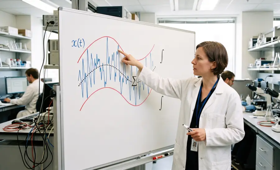
Empirical mode decomposition (EMD) was developed as a response to a mismatch between the EEG processing tools and the data it is being asked to interpret. Rather than forcing a signal into a fixed set of sine waves or wavelets chosen in advance, EMD lets the data define its own building blocks. These building blocks are called intrinsic mode functions (IMFs), and the process that generates them requires no assumption that the signal is stationary or linear.
This makes EMD conceptually appealing for EEG, where transient, irregular events are often the exact features a researcher wants to detect. That appeal has driven a body of applied research into seizure detection, emotion classification, artifact removal, and brain-computer interfacing.
Why EEG Signals Resist Traditional Signal Processing Tools
The core problem with applying classic signal processing methods to EEG is a structural one. Fourier-based analysis decomposes a signal into a fixed library of sine and cosine waves, an approach that works cleanly when the underlying process does not change its statistical behavior over time.
EEG does not meet that condition. A person's brain activity during quiet rest looks statistically different from the same person's activity during a burst of focused attention or, in a clinical setting, during a seizure. Research cites this non-stationary quality as the starting justification for turning to EMD instead of frequency-based tools built for stable signals.
The practical consequence is that a method assuming stationarity can smear or misrepresent transient neural events, treating a brief spike as if it were spread evenly across a longer window. For applications like epilepsy diagnosis, where the timing and shape of a spike carries diagnostic weight, that kind of smearing is a real limitation.
EMD's proposed solution is to avoid the fixed-basis assumption entirely and let the decomposition adapt to whatever shape the signal actually takes.
What Empirical Mode Decomposition Does and How It Works
EMD operates through a repeated process called sifting. The algorithm identifies the local peaks and troughs of a signal, connects them into upper and lower envelopes, and subtracts the average of those envelopes from the original signal.
This step repeats until the resulting component meets two conditions:
Its upper and lower envelopes are symmetric around zero
Its mean stays close to zero across the whole segment.
Once a component satisfies these conditions, it is labeled an intrinsic mode function (IMFs), and it gets subtracted from the original signal so the process can extract the next one.
The output is a stack of IMFs ordered from the fastest oscillations to the slowest, followed by a final leftover trend that captures whatever slow drift remains. Because each IMF is extracted directly from the local shape of the data rather than matched against a predetermined waveform, EMD can, in principle, represent irregular and shifting dynamics without needing the analyst to pick a basis in advance.
Method | Best Condition |
|---|---|
EMD | Heavy contamination |
ICA or CCA | Light noise, single source |
Wavelet Transform | Light noise, two sources |
How EMD Captures Transient Events Like Seizure Spikes
Transient events in EEG, epileptiform spikes, or short bursts tied to a cognitive task, carry outsized clinical and neuroscientific importance, but they are often buried under background brain activity or contaminating noise. This is precisely the scenario where EMD's local, adaptive decomposition is meant to offer an edge over fixed-basis methods.
A study by Safieddine et al. tested this directly by simulating epileptic spike-like signals and contaminating them with real muscle artifact at different intensities, then comparing four correction methods:
EMD
Independent component analysis (ICA)
Canonical correlation analysis (CCA)
Wavelet transform
When contamination was heavy, EMD outperformed all three alternatives. That advantage was not absolute, however. When the signal was less noisy and the spikes came from a single cortical source, CCA and ICA produced better results. When spikes originated from two distinct sources, either EMD or ICA worked best under heavy noise, while wavelet transform performed better under lighter noise.
In other words, the best artifact-removal tool depended on both the noise level and the underlying source configuration, and EMD's edge was strongest specifically in the heavily contaminated case.
Meanwhile, research studies by Riaz et al. and Shafiul Alam et al. moved from artifact correction to classification, using IMFs as a source of features for detecting seizures and epilepsy.
Riaz’s team extracted temporal moments, meaning statistical measures like variance, skewness, and kurtosis, along with spectral features such as spectral centroid and spectral skew, from each IMF. These features were fed into a support vector machine, a standard supervised learning classifier, and the resulting classification of epilepsy patients and seizure segments produced what the authors described as good results that compared favorably against other established feature-extraction methods.
Alam’s team pushed further using higher-order statistical moments calculated within the EMD domain, feeding them into an artificial neural network. Across several classification tasks, including distinguishing seizure activity from non-seizure activity, the method reported accuracy, sensitivity, and specificity as high as 100 percent, while running faster than comparable time-frequency analysis techniques.
Taken together, these studies suggest EMD can help isolate transient epileptic activity, but its performance is sensitive to noise level, source geometry, and the dataset it is tested on rather than being a fixed, guaranteed advantage.
Common Challenges in Empirical Mode Decomposition
EMD is flexible, but that flexibility creates methodological choices that can affect scientific conclusions. Small perturbations in a signal may change the extrema, and therefore change the envelopes and extracted modes. Noise, filtering, short recordings, and sharp boundaries can all make the decomposition less stable.
Common checks help expose rather than conceal these difficulties. They include:
Measuring reconstruction error after summing the IMFs and residual.
Testing sensitivity to endpoint treatment and sifting thresholds.
Comparing decompositions across repeated segments or noise ensembles.
Inspecting whether a removed component contains plausible neural activity.
These checks do not eliminate ambiguity, but they make it visible. Mode mixing may call for an ensemble variant, while excessive high-frequency content may indicate contamination rather than a useful neural mode. In all cases, the analysis should report the decisions that produced the final representation.
Another challenge is communication. An IMF index is an algorithmic label, not a diagnosis or a universally defined EEG band. Clear figures, parameter records, and cautious language are needed when results move from exploratory signal processing into biomedical interpretation.
Mode Mixing: The Main Weakness of Basic EMD
The clearest structural weakness in basic EMD is a phenomenon called mode mixing. It happens in two related ways. Either oscillations that operate on very different time scales end up trapped inside the same IMF, or a single, coherent rhythmic component gets fragmented and scattered across several IMFs instead of staying contained in one. Both versions of the problem make it harder to trust that an IMF corresponds to a genuine, singular physiological process.
The practical risk is interpretive. If a researcher assumes a given IMF represents, say, activity in a specific frequency band, but that IMF actually contains a blend of muscle artifact and residual spike activity due to mode mixing, any conclusion drawn from that IMF becomes unreliable.
Complete Ensemble Empirical Mode Decomposition
Complete ensemble empirical mode decomposition, commonly called CEEMD, is a noise-assisted extension of EMD. It adds carefully controlled noise realizations to the signal, decomposes the resulting ensemble, and averages corresponding modes. The aim is to make the decomposition more stable when nearby oscillatory scales are difficult to separate.
How CEEMD Improves on Standard EMD
Standard EMD can suffer from mode mixing, in which oscillations with different scales appear in one IMF or similar scales are spread across several IMFs. CEEMD addresses this instability statistically rather than changing the basic envelope-sifting idea. Repeated decompositions and averaging can reduce the influence of an individual noise realization, but the outcome still depends on the ensemble design, noise amplitude, number of trials, and implementation details.
The method is not automatically superior for every dataset. It increases computational cost and can retain residual noise or introduce boundary sensitivity if its parameters are poorly chosen. For EEG, comparisons should therefore examine reconstruction quality, stability across trials, preservation of transient events, and whether the resulting modes remain interpretable for the stated research question.
What EEG Applications Have Tested EMD So Far
EMD has been commonly applied in three broad categories.
Feature extraction for classification represents the largest use case. For instance, a study led by Ning Zhuang used EMD for emotion recognition, extracting multidimensional features from IMFs, including the first difference of the time series, the first difference of phase, and normalized energy, then testing them on the publicly available DEAP emotional database. The study found the high-frequency IMF1 component had a significant effect on distinguishing emotional states, and the overall approach outperformed comparison methods including fractal dimension, sample entropy, and discrete wavelet transform.
Artifact removal is represented in studies like the one by Safieddine et al., where EMD's deterministic, envelope-based decomposition removed muscle artifact from simulated epileptic EEG more effectively than ICA, CCA, or wavelet-based methods, specifically under conditions of heavy contamination.
Lastly, multichannel analysis and brain-computer interfacing can be seen in research published in the Journal of Circuits, Systems and Computers, where EMD was applied separately to each channel in a multichannel EEG recording, and the resulting IMFs were clustered according to spectral similarity to identify which components correlated with an external stimulus.
Reconstructing the signal from only those stimulus-linked components allowed the researchers to identify when a subject was attending to a given stimulus, a capability directly relevant to BCI systems that depend on distinguishing intentional neural signals from background noise.
Empirical Mode Decomposition Tutorial for Beginners
A beginner can think of EMD as repeatedly peeling the quickest visible oscillation away from a complicated waveform. The algorithm does not begin with a list of frequency bands. Instead, it observes where the signal rises and falls, estimates a local centerline, and removes that centerline from the candidate oscillation.
A sensible learning exercise starts with a short, clean synthetic signal containing two oscillations and a slow trend. The expected result is not a perfect separation in every case, but a set of components whose sum approximately reproduces the input. Plotting the original signal, every IMF, and the residual on a shared time axis is often more informative than inspecting numerical output alone.
For an EEG recording, the workflow also includes decisions that precede decomposition:
Selecting channels and segments
Checking sampling information
Marking bad intervals
Documenting preprocessing
A general frequency-decomposition guide can help situate EMD among Fourier, wavelet, and related time–frequency methods. These choices do not turn EMD into a clinical test; they define the conditions under which its output can be evaluated.
Empirical Mode Decomposition Python: A Coding Walkthrough
Python implementations typically accept a one-dimensional array and return a collection of IMFs, sometimes with a residual or through a separate residue operation. A basic workflow loads a signal, confirms its sampling order and missing-value status, runs the decomposition, and plots the components. Reproducible analysis also records the library version and the parameters controlling sifting and ensemble behavior.
Popular Python Libraries for EMD
Several scientific Python packages provide EMD or related algorithms, but their APIs and defaults differ. Some expose standard EMD, ensemble variants, stopping thresholds, spline choices, and visualization helpers; others require more manual assembly. The central coding task is not merely calling a function, but checking what the returned components mean and whether their sum reconstructs the input.
A minimal pseudocode pattern is:
The code is intentionally schematic because function names and return values vary across packages. Validation should include a reconstruction error, plots of extrema and boundaries, and sensitivity checks under reasonable parameter changes.
For EEG, data dimensions also matter: a multichannel recording may require a channel-wise workflow or a specifically multivariate method rather than silently flattening all channels into one array.
Applying Empirical Mode Decomposition to EEG Signals
EEG is a natural setting for empirical mode decomposition because voltage changes can contain overlapping rhythms, transient events, and non-stationary activity. EMD may be applied to individual channels or selected segments to examine how local oscillations evolve. The relevant scientific question might concern temporal structure, artifact separation, event-related changes, or features for a subsequent statistical model.
Before decomposition, signal quality and recording context need attention. Electrode placement, reference scheme, sampling rate, filtering, missing samples, and marked artifacts can all influence local extrema.
Moreover, the resulting IMFs should not be assigned physiological labels solely by their order. Fast components may include muscle or instrument noise, and slower components may contain drift as well as meaningful activity.
Interpretation is stronger when it compares reconstructed signals, retains a clear audit trail of excluded components, and tests whether findings persist across channels, participants, and analysis choices.
Empirical Mode Decomposition vs Wavelet Transform
EMD and the wavelet transform both support time–frequency analysis, but they organize signal structure differently. EMD derives modes from local extrema in the observed data, whereas wavelet analysis compares the signal with a selected family of scaled and shifted basis functions. One is adaptive to the data; the other provides a predefined multiscale coordinate system.
The distinction becomes clearer when the analytical priorities are compared:
Feature | Empirical mode decomposition | Wavelet transform |
|---|---|---|
Representation | Data-derived intrinsic mode functions | Coefficients on chosen wavelet scales |
Basis | Adaptive and signal dependent | Prespecified wavelet family |
Time localization | Local, with data-dependent modes | Scale-dependent and controlled by the wavelet |
Main concerns | Mode mixing, endpoint effects, stopping rules | Basis choice, resolution trade-offs, boundary effects |
Neither method is universally preferable. EMD can offer an intuitive decomposition for changing oscillations, while wavelets can provide a consistent framework for comparing signals when the basis and scales are scientifically appropriate.
The choice should follow the question, the expected transients, the need for reproducibility, and the validation strategy rather than a general claim that one method is more advanced.
What Brain Signal Analysis Gains from Data-Driven Decomposition
Brain activity rarely holds still long enough for classic frequency tools to catch its most telling moments, which is why a method that lets the signals define their own building blocks has drawn real attention.
Empirical mode decomposition steps around the need for fixed mathematical templates, producing results that have proven useful for isolating seizure spikes under heavy noise and extracting meaningful features for emotion recognition. This shift toward adaptively shaped analysis creates a pathway for catching transient neural events that would otherwise be smeared or missed. The practical outcome, as seen across the studies, is a tool that can aid classification and artifact removal when conditions align in its favor.
References
Safieddine, D., Kachenoura, A., Albera, L., Birot, G., Karfoul, A., Pasnicu, A., ... & Merlet, I. (2012). Removal of muscle artifact from EEG data: comparison between stochastic (ICA and CCA) and deterministic (EMD and wavelet-based) approaches. EURASIP Journal on Advances in Signal Processing, 2012(1), 127. https://doi.org/10.1186/1687-6180-2012-127
Riaz, F., Hassan, A., Rehman, S., Niazi, I. K., & Dremstrup, K. (2015). EMD-based temporal and spectral features for the classification of EEG signals using supervised learning. IEEE Transactions on Neural Systems and Rehabilitation Engineering, 24(1), 28-35. https://doi.org/10.1109/TNSRE.2015.2441835
Alam, S. S., & Bhuiyan, M. I. H. (2013). Detection of seizure and epilepsy using higher order statistics in the EMD domain. IEEE journal of biomedical and health informatics, 17(2), 312-318. https://doi.org/10.1109/JBHI.2012.2237409
Zhuang, N., Zeng, Y., Tong, L., Zhang, C., Zhang, H., & Yan, B. (2017). Emotion recognition from EEG signals using multidimensional information in EMD domain. BioMed research international, 2017(1), 8317357. https://doi.org/10.1155/2017/8317357
Rutkowski, T. M., Mandic, D. P., Cichocki, A., & Przybyszewski, A. W. (2010). EMD approach to multichannel EEG data—the amplitude and phase components clustering analysis. Journal of Circuits, Systems, and Computers, 19(01), 215-229. https://doi.org/10.1142/S0218126610006037
Frequently Asked Questions
What is empirical mode decomposition (EMD) and why is it useful for EEG?
EMD is an adaptive signal processing method that breaks down a waveform into oscillatory components called intrinsic mode functions without assuming the signal is stationary. It is useful for EEG because brain activity shifts continuously and contains transient events, which traditional fixed-basis tools struggle to represent accurately.
How does the sifting process in EMD work?
The sifting process repeatedly identifies a signal's local peaks and troughs, constructs upper and lower envelopes, and subtracts their average from the signal. This step repeats until the remaining component has symmetric envelopes and a near-zero mean, at which point it becomes an intrinsic mode function.
What are intrinsic mode functions (IMFs)?
IMFs are the data‑driven building blocks produced by EMD, ordered from the fastest oscillations to the slowest. Because they are extracted directly from the local shape of the signal rather than matched to a preset waveform, they can capture irregular and shifting neural dynamics.
Why is mode mixing a problem in EMD?
Mode mixing occurs when oscillations of different time scales become trapped in the same IMF or when a single rhythm gets split across multiple IMFs. This ambiguity makes it hard to trust that an IMF represents a single physiological process, potentially blending noise or artifacts with genuine brain activity.
How does EMD handle transient events like seizure spikes?
EMD isolates brief spikes by extracting them into high‑frequency IMFs, where background activity is separated. Under heavy muscle contamination, EMD outperformed other artifact‑removal methods, though its advantage diminished with cleaner signals or different source configurations.
Can EMD replace traditional methods like the Fourier transform for all EEG analysis?
EMD is not a universal replacement; it is most valuable for non-stationary and transient signals where fixed‑basis Fourier methods fall short. Its reliability, however, varies with noise levels and is not yet proven across all EEG recording conditions.
Does EMD require assuming the signal is stationary?
No, EMD does not assume stationarity, a key advantage over classical Fourier analysis for EEG. The decomposition adapts directly to the data’s changing statistical character, allowing it to track shifting brain dynamics and brief events without preconditioning.
Can EMD analyze multichannel EEG directly?
Basic EMD is commonly applied to one-dimensional signals. Multichannel analysis requires a deliberate channel-wise or multivariate strategy, because simply combining channels can obscure spatial and temporal meaning.
Emotiv is a neurotechnology leader helping advance neuroscience research through accessible EEG and brain data tools.
Medical Disclaimer: The information provided on this website is for educational and informational purposes only and is not intended as medical or health advice. This content may contain errors and should not be relied upon to make life-altering health, medical, or lifestyle choices. Always seek the advice of your physician or other qualified health provider with any questions you may have regarding a medical condition or treatment.
Christian Burgos


