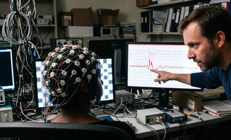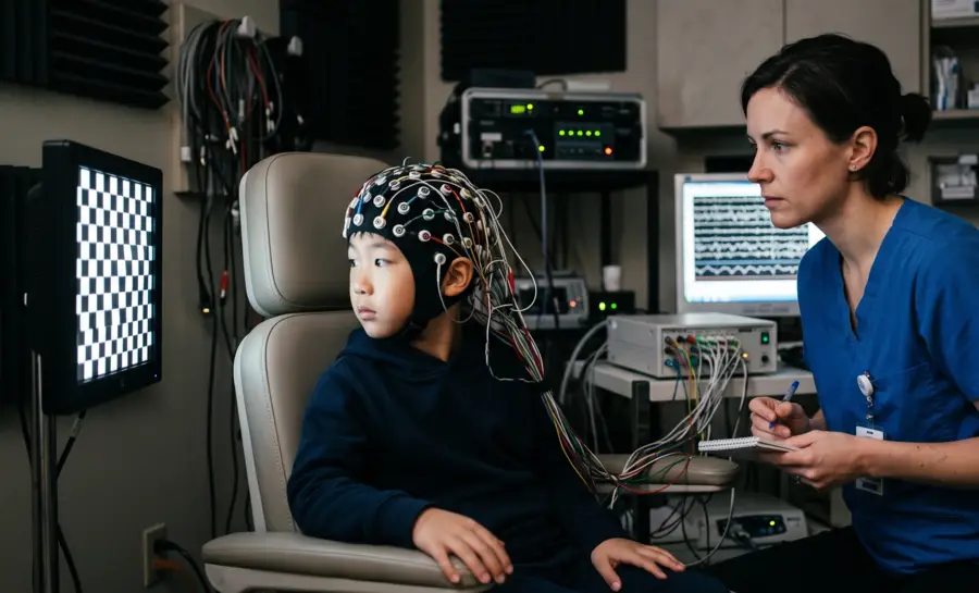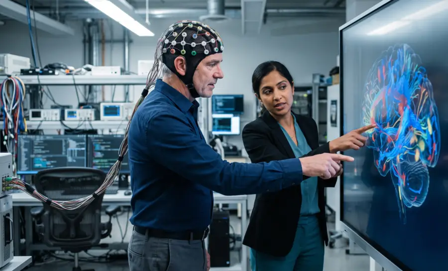
A cortical evoked potential (CEP) is the immediate, time-locked voltage change that occurs when a population of cortical neurons is perturbed directly, usually with a transcranial magnetic stimulation (TMS) pulse or a brief electrical current applied to the cortical surface.
The signal appears on an electroencephalogram (EEG) as a small, fast deflection, but its defining feature is its origin. The cortical label signals that the recorded activity reflects local excitability, synaptic processing within the cortex, and transmission between cortical areas, rather than the arrival of sensory information from the eyes, ears, or a subcortical relay station.
What are Cortical Evoked Potentials?
Cortical evoked potentials are measurable changes in electrical activity that follow a defined sensory or electrical stimulus. Unlike spontaneous EEG activity, they are time-locked to an event and are assessed by examining features such as:
Latency
Amplitude
Waveform shape
Spatial distribution
These responses provide a window into the timing and integrity of neural pathways that connect the periphery with the cortex.
The Role of the Cortex in Evoked Potentials
The cortex is the destination for many sensory pathways, but cortical responses do not occur in isolation. Signals are filtered and transformed as they pass through peripheral receptors, cranial or spinal pathways, the brainstem, and thalamic circuits. The final response therefore reflects both pathway conduction and the cortical networks that interpret or integrate the incoming information.
Cortical evoked potentials may also vary with alertness, attention, age, medications, and the condition of the nervous system. Their timing can offer information about transmission speed, while their distribution can suggest which cortical regions participate in the response. For broader background on stimulus-locked activity and its distinction from spontaneous brain signals, see this overview of evoked potentials in EEG.
Where do CEP Signals Originate in the Cortex?
The EEG signal that forms a CEP emerges from the summed postsynaptic potentials of tens of thousands of pyramidal neurons arranged in parallel within the cortex.
When a TMS pulse or cortical electrical stimulus drives a cortical population, synapses release neurotransmitters, postsynaptic receptors open, ions flow, and the resulting postsynaptic currents create a measurable electrical field. The CEP is the EEG readout of those intrinsic cortical currents.
A useful example comes from TMS-evoked cortical potentials in the motor cortex. When a single TMS pulse activates the cortex, it produces a sequence of waveform components.
An early negative potential appears around 45 milliseconds after the pulse. A later negative potential emerges around 100 milliseconds.
Each component carries distinct physiological signatures because different cortical synapses and receptor systems shape them. The early component and the late component respond differently to drugs that manipulate GABA-A and GABA-B receptors, as the pharmaco-TMS-EEG experiments show. That pharmacological separation tells us the waveform components are layered readouts of specific postsynaptic circuitry.
How to Distinguish Peripheral vs. Cortical TMS Responses
A TMS pulse delivered to the scalp is not a clean, cortex-only intervention. The magnetic field also excites sensory axons in the scalp, triggers small muscle twitches near the coil, and generates a sharp clicking sound.
That click activates the auditory cortex, and the scalp sensation activates somatosensory cortex. Those secondary sensory inputs produce cortical potentials with a temporal and spatial pattern that can closely resemble the CEPs produced by direct cortical stimulation.
As a consequence, a realistic sham control tries to address this by mimicking the sound and scalp sensation of TMS while avoiding transcranial cortical stimulation. In a 2018 study, when researchers delivered sham TMS with the coil oriented to reduce direct cortical activation, the sham-induced potentials overlapped strongly with the real TMS potentials. This overlap persisted even when investigators used foam padding under the coil and auditory noise masking to attenuate the peripheral confounds.
Moreover, the authors reported that in 17 healthy young volunteers, the temporal and spatial features of early and late TMS-evoked potential components after real TMS closely resembled the potentials after realistic sham TMS, whether tested over the posterior parietal cortex or the superior frontal gyrus.
These findings underscore how difficult it is to separate genuine cortical responses from peripheral sensory confounds. Without rigorous control conditions, distinguishing direct neural excitation from secondary sensory feedback remains a significant hurdle.
How CEPs Trace Cortico-Cortical Connectivity
When researchers record from subdural electrode grids placed directly on the cortical surface, a different version of the CEP emerges: the cortico-cortical evoked potential.
Here, a small electrical pulse is delivered to one cortical region and the resulting activity is recorded at other cortical electrodes. Because both stimulation and recording occur within the cortex, peripheral sensory confounds from the scalp are eliminated. This setup turns CEPs into connectivity tracers, mapping the pathways that link cortical regions.
Evidence from the human language system illustrates how this works. In eight patients undergoing invasive epilepsy monitoring, researchers first used conventional cortical electrical stimulation to identify the anterior and posterior language areas. They then delivered single electrical pulses to those areas and averaged the resulting electrocorticograms from perisylvian and extrasylvian basal temporal language electrodes.
Therein, the researchers reported that stimulation of the anterior language area produced cortico-cortical evoked potentials in the lateral temporo-parietal area in seven of eight patients. The responses appeared in the middle and posterior parts of the superior temporal gyrus, the adjacent middle temporal gyrus, and the supramarginal gyrus.
Importantly, the potentials also occurred at or near the specific posterior language electrodes that produced speech arrest when stimulated. Stimulation of the adjacent face motor area, by contrast, did not elicit responses in language areas. That response appeared in the postcentral gyrus instead.
What do these propagated potentials tell us about neural generators?
Each so-called late evoked potential appears at a cortical electrode only after a coupling delay. That delay reflects the time needed for neuronal activity to travel along cortico-cortical pathways from the stimulation site to the recording site. Therefore, the recorded potential appeared to represent neuronal activity transmitted over anatomical connections between cortical regions.
The language data also showed a more networked architecture than the classical Wernicke-Geschwind model proposed. Stimulation of the anterior language area produced potentials in the posterior language region and basal temporal areas. Meanwhile, stimulation of the posterior language area produced potentials in the anterior language area and, in a subset of patients, the basal temporal area.
This bidirectional pattern suggests feed-forward and feed-back projections between Broca's and Wernicke's regions, potentially mediated by the arcuate fasciculus, the cortico-subcortico-cortical pathway, or both.
Furthermore, the potentials were recorded from a larger cortical territory than the posterior language area identified by electrical stimulation alone, suggesting a broader neuronal network surrounds the recognized core region.
Neurotransmitter Signatures of Cortical Evoked Potentials
The TMS-evoked EEG potential’s components shift predictably when specific cortical synapses are manipulated with drugs. The cortex seems to rely on GABAergic inhibition to balance excitation.
GABA-A receptors mediate fast inhibitory signaling. GABA-B receptors mediate slower, longer-lasting inhibition. The aforementioned pharmaco-TMS-EEG experiments tested the effects of compounds acting on each system, focusing on the N45 component around 45 milliseconds and the N100 component around 100 milliseconds.
The findings followed a consistent pattern:
Positive modulation of GABA-A receptors with benzodiazepines, specifically alprazolam and diazepam, increased the amplitude of the early negative component at approximately 45 milliseconds.
Zolpidem, which acts primarily on α1-subunit-containing GABA-A receptors, also increased the N45, but left the later component unchanged.
Benzodiazepines tended to reduce the late negative component by around 100 milliseconds.
Baclofen, a drug that directly activates GABA-B receptors, specifically increased the N100 amplitude.
These differential effects suggest that the N45 component appears to represent activity driven by α1-subunit-containing GABA-A receptors. The N100 component appears to represent GABA-B receptor activity. It is like the waveform’s components map onto specific synaptic receptor mechanisms in the human cortex.
That makes TMS-EEG components candidate markers for studying inhibitory and excitatory balance in conditions such as epilepsy or schizophrenia, where GABAergic signaling is implicated.
How CEPs Reflect Brain State and Age-Dependent Changes
Cortical evoked potential amplitude is not fixed even within the same individual. It shifts with the intrinsic state of the cortex at the moment of stimulation.
A sleep slow-oscillation study tried to demo this directly. When researchers targeted the primary motor cortex with single-pulse TMS during non-rapid eye movement sleep, they triggered stimulation online based on automatic EEG detection of slow oscillation up-states and down-states.
Up-states correspond to phases of global depolarization. Down-states correspond to phases of hyperpolarization.
The authors found that compared to wakefulness, sleep motor-evoked potentials were smaller and delayed. Sleep TMS-evoked EEG potentials were fundamentally altered, closely resembling a spontaneous slow oscillation. But the critical state-dependent finding was that both motor-evoked potentials and TMS-evoked EEG potentials were consistently larger when evoked during up-states than during down-states.
Furthermore, amplitudes within each sleep state depended on the actual EEG potential at the exact time and site of stimulation. The cortex’s excitability shifts within the oscillation itself, with millisecond resolution. This finding shows that brain wave phase and cortical excitability are coupled in a measurable way.
A parallel story emerges from aging research, but with a sharp distinction between physiological and pathological change. When researchers compared TMS-evoked EEG potentials in healthy elderly individuals, healthy young individuals, and patients with Alzheimer's disease, they found that physiological aging did not alter the EEG response to TMS of the left superior frontal cortex. The healthy elderly and the healthy young produced similar cortical potentials.
The response was markedly altered only in patients with Alzheimer's disease and cognitive impairment. This dissociation suggests the CEP is not sensitive to normal age-related cortical changes, but is sensitive to pathological cortical dysfunction associated with cognitive decline.
Across sleep and aging, the same principle holds: CEPs rely on cortical membrane potentials, synaptic state, and the brain state present at the time of stimulation. Because of that dependence, they allow neuroscientists to probe changes in cortical excitability over time, across states, and in relation to pathological processes.
Future Directions and Research in Cortical Evoked Potentials
Research is moving toward more precise descriptions of how evoked responses vary across individuals, trials, cortical regions, and states of consciousness.
High-density scalp recordings, improved intracranial sampling, and combined anatomical-functional mapping may help clarify the relationship between recorded voltage and underlying neural sources. Better standards for stimulus delivery and metadata are equally important because technical consistency supports meaningful comparison.
Signal analysis is also expanding beyond simple visual identification of peaks. Single-trial methods, statistical modeling, source estimation, and carefully validated machine-learning approaches may preserve information that conventional averaging removes. These methods must still be tested against artifact sensitivity, dataset shift, interpretability, and clinically meaningful endpoints rather than judged only by predictive performance.
Lastly, future studies will benefit from transparent reporting and shared analytical definitions. Common descriptions of stimulation, recording montages, preprocessing, response windows, and significance criteria could make findings easier to reproduce across laboratories. The broader goal is not merely to obtain larger waveforms, but to connect cortical evoked potentials with reliable measures of pathway function, network communication, and patient-relevant physiology.
Why Direct Cortical Listening Changes How We Study Brain Health
Cortical evoked potentials reframe the question of how we hear the brain: instead of tracking sensory signals from the outside world, they capture the cortex's own response to direct stimulation.
These signals reflect local synaptic machinery, receptor-specific activity, and communication between cortical regions, all of which shift with brain state and disease. By linking specific waveform components to GABAergic receptor systems and mapping bidirectional language pathways, the technique turns an EEG deflection into a functional readout of cortical physiology.
Noteworthy, that readout only becomes trustworthy when researchers separate the cortex from the scalp and ear. Realistic sham controls and invasive recordings show that direct cortical activation can be distinguished from peripheral sensory noise, and the same controls make drug studies and aging comparisons interpretable.
As a result, cortical evoked potentials are best understood as practical markers for measuring cortical excitability, connectivity, and pathological dysfunction in conditions like epilepsy, schizophrenia, and Alzheimer's disease, rather than as fixed answers about how the brain works.
References
Premoli, I., Castellanos, N., Rivolta, D., Belardinelli, P., Bajo, R., Zipser, C., ... & Ziemann, U. (2014). TMS-EEG signatures of GABAergic neurotransmission in the human cortex. The Journal of Neuroscience, 34(16), 5603-5612. https://doi.org/10.1523/JNEUROSCI.5089-13.2014
Conde, V., Tomasevic, L., Akopian, I., Stanek, K., Saturnino, G. B., Thielscher, A., ... & Siebner, H. R. (2019). The non-transcranial TMS-evoked potential is an inherent source of ambiguity in TMS-EEG studies. Neuroimage, 185, 300-312. https://doi.org/10.1016/j.neuroimage.2018.10.052
Matsumoto, R., Nair, D. R., LaPresto, E., Najm, I., Bingaman, W., Shibasaki, H., & Lüders, H. O. (2004). Functional connectivity in the human language system: a cortico-cortical evoked potential study. Brain, 127(10), 2316-2330. https://doi.org/10.1093/brain/awh246
Bergmann, T. O., Mölle, M., Schmidt, M. A., Lindner, C., Marshall, L., Born, J., & Siebner, H. R. (2012). EEG-guided transcranial magnetic stimulation reveals rapid shifts in motor cortical excitability during the human sleep slow oscillation. The Journal of Neuroscience, 32(1), 243-253. https://doi.org/10.1523/JNEUROSCI.4792-11.2012
Casarotto, S., Määttä, S., Herukka, S. K., Pigorini, A., Napolitani, M., Gosseries, O., ... & Massimini, M. (2011). Transcranial magnetic stimulation-evoked EEG/cortical potentials in physiological and pathological aging. Neuroreport, 22(12), 592-597. https://doi.org/10.1097/WNR.0b013e328349433a
Frequently Asked Questions
What is a cortical evoked potential?
A cortical evoked potential is a time-locked voltage change recorded on an EEG that occurs when a population of cortical neurons is directly stimulated, usually with a TMS pulse or electrical current. Unlike responses to sensory events, it reflects the cortex's internal excitability and synaptic processing rather than incoming sensory information.
How does a cortical evoked potential differ from a sensory evoked potential?
A sensory evoked potential tracks how an external stimulus travels from a sense organ through the brainstem and thalamus into the cortex, while a cortical evoked potential begins with direct stimulation of the cortex itself. The difference is one of direction: sensory potentials trace incoming (afferent) input, while cortical evoked potentials probe local and outgoing processing within the cortical tissue.
Where do the signals for a cortical evoked potential come from?
The signals come from the summed postsynaptic potentials of tens of thousands of pyramidal neurons arranged in parallel within the cortex. When a direct pulse drives these neurons, synapses release neurotransmitters and ions flow, creating a measurable electrical field that becomes the evoked potential.
Why is a sham control necessary when studying TMS-evoked potentials?
A TMS pulse also excites sensory axons in the scalp, triggers muscle twitches, and creates a clicking sound that activates auditory and somatosensory cortex, producing potentials that resemble direct cortical responses. A realistic sham control mimics these peripheral sensations while avoiding transcranial stimulation, allowing researchers to separate true cortical activation from peripheral contributions. Without that control, the EEG response could reflect peripheral sensory processing rather than direct cortical activation.
How do different drugs affect the waveform components of cortical evoked potentials?
Drugs that positively modulate GABA-A receptors increase the early negative component of the TMS-evoked potential, while zolpidem specifically affects that early component without changing the later one. Baclofen, which activates GABA-B receptors, increases the late negative component. This shows that different waveform components map onto specific postsynaptic receptor mechanisms in the cortex.
How do cortical evoked potentials change with brain state and aging?
Cortical excitability shifts within a brain oscillation itself, with millisecond resolution, meaning the amplitude of the evoked potential depends on the phase of the brain wave at the time of stimulation. Physiological aging does not alter the cortical response to TMS, but pathological conditions like Alzheimer's disease produce markedly different potentials, suggesting the CEP is sensitive to pathological cortical dysfunction but not normal age-related changes.
What does stimulation of language areas reveal about cortical connectivity?
Direct stimulation of the anterior language area produces evoked potentials in posterior language regions and basal temporal areas, while stimulation of the posterior language area produces responses in the anterior language area. This bidirectional pattern suggests feed-forward and feed-back projections between Broca's and Wernicke's regions, revealing a broader networked architecture than the classical model proposed.
What are cortico-cortical evoked potentials, and how are they used?
Cortico-cortical evoked potentials are recorded by delivering a small electrical pulse to one cortical region and measuring the response from neighboring cortical sites using subdural electrode grids placed on the brain's surface. This setup eliminates peripheral confounds and allows scientists to map the anatomical pathways that link different cortical regions.
Emotiv is a neurotechnology leader helping advance neuroscience research through accessible EEG and brain data tools.
Medical Disclaimer: The information provided on this website is for educational and informational purposes only and is not intended as medical or health advice. This content may contain errors and should not be relied upon to make life-altering health, medical, or lifestyle choices. Always seek the advice of your physician or other qualified health provider with any questions you may have regarding a medical condition or treatment.
Christian Burgos