Bulbar-onset patients experience more rapid functional decline, earlier respiratory compromise, and higher rates of cognitive impairment. Statistical analyses consistently demonstrate that bulbar-onset ALS correlates with accelerated disease progression and reduced survival times.
What Is the Neuroanatomical Basis for Bulbar Symptoms in ALS?
The temporal sequence of bulbar symptom development follows predictable anatomical patterns.
Speech articulation typically deteriorates first, reflecting the high precision demands placed on coordinated movements of the tongue, lips, and soft palate. Swallowing difficulties often follow, beginning with liquids before progressing to solid foods.
This progression reflects the complex neuromuscular coordination required for safe deglutition, involving multiple cranial nerve nuclei working in precise temporal sequences.
Trigeminal motor nucleus (CN V) controls mastication, with early involvement causing difficulty in food bolus preparation.
Facial nucleus (CN VII) innervates facial expression muscles and maintains lip seal during swallowing and speech.
Glossopharyngeal (CN IX) and vagus (CN X) nuclei coordinate swallowing reflexes and contribute to soft palate elevation and voice quality.
Hypoglossal nucleus (CN XII) drives intrinsic and extrinsic tongue muscles, with degeneration leading to visible atrophy and fasciculations.
Accessory nucleus (CN XI) supplies sternocleidomastoid and trapezius muscles, contributing to neck weakness and head drop.
Cranial Nerve | Key Function |
|---|---|
V (Trigeminal) | Chewing muscles |
VII (Facial) | Facial expression, lip seal |
IX, X (Glossopharyngeal/Vagus) | Swallowing, voice, airway |
XII (Hypoglossal) | Tongue movement, speech |
XI (Accessory) | Head and neck strength |
How Does Degeneration of Corticobulbar Tracts Contribute to Bulbar ALS Symptoms?
Upper motor neuron degeneration within corticobulbar pathways produces spastic dysarthria, characterized by slow, labored speech with a strained-strangled voice quality. This contrasts with the flaccid dysarthria resulting from lower motor neuron involvement, which produces breathy, weak vocalization. Many bulbar-onset patients demonstrate mixed dysarthria, reflecting combined upper and lower motor neuron pathology.
Corticobulbar tract degeneration also underlies pseudobulbar affect, the inappropriate and involuntary episodes of laughing or crying that affect many ALS patients. This phenomenon reflects loss of cortical inhibition over brainstem emotional expression circuits.
While distressing for patients and families, pseudobulbar affect serves as an important clinical marker of upper motor neuron involvement in bulbar territories.
The bilateral nature of most corticobulbar innervation initially provides functional compensation when unilateral pathology develops. However, this same redundancy can mask early disease progression, leading to delayed recognition of bulbar involvement.
Once bilateral pathology reaches critical thresholds, functional decline accelerates rapidly, explaining the characteristic steep progression curves observed in bulbar-onset patients.
Corticobulbar pathology also affects respiratory control circuits in the brainstem, contributing to the early respiratory compromise observed in many bulbar-onset patients.
Why Is the Hypoglossal Nucleus Particularly Vulnerable in Bulbar ALS?
The hypoglossal nucleus demonstrates selective vulnerability in ALS that exceeds even other cranial nerve nuclei. This preferential targeting reflects several unique anatomical and physiological characteristics.
Hypoglossal motor neurons are among the largest in the brainstem, with extensive dendritic trees and high metabolic demands. These features parallel the selective vulnerability of large spinal motor neurons in limb-onset ALS.
Hypoglossal motor neurons exhibit unique calcium handling properties that may contribute to their vulnerability. These neurons rely heavily on calcium-binding proteins to manage intracellular calcium homeostasis during high-frequency firing.
Disruption of calcium homeostasis represents a key pathogenic mechanism in ALS, and hypoglossal neurons may be particularly susceptible to calcium-mediated excitotoxicity.
The hypoglossal nucleus also receives convergent inputs from multiple cortical and subcortical regions involved in speech, swallowing, and respiratory control. This extensive connectivity may facilitate the spread of pathological proteins or other toxic factors from affected regions.
What Do Genetic and Pathological Findings Reveal About Bulbar-Onset ALS?
The genetics of bulbar-onset ALS reveals distinct patterns that distinguish it from limb-onset disease. These genetic associations provide crucial insights into the biological mechanisms underlying bulbar vulnerability and help explain the more aggressive disease course characteristic of this presentation.
Pathological studies demonstrate that bulbar-onset ALS exhibits unique patterns of protein aggregation and cellular degeneration. The distribution and characteristics of pathological protein deposits differ between bulbar and limb-onset cases, suggesting distinct pathogenic pathways.
Moreover, the strong association between bulbar-onset ALS and cognitive impairment reflects shared genetic and pathological features with frontotemporal dementia.
Understanding these connections provides insights into the broader spectrum of neuroscience underlying motor neuron diseases.
How Do C9orf72 Gene Expansions Correlate with Bulbar Onset?
The C9orf72 gene contains a hexanucleotide repeat sequence (GGGGCC) that, when abnormally expanded, represents the most common known genetic cause of ALS.
Patients carrying pathological C9orf72 expansions demonstrate a significantly higher likelihood of presenting with bulbar symptoms compared to other genetic or sporadic forms of ALS.
This genetic association extends beyond simple onset patterns. C9orf72 expansion carriers with bulbar onset demonstrate more rapid disease progression and shorter survival times compared to C9orf72 carriers with limb onset. The mechanism underlying this association likely involves the preferential vulnerability of brainstem motor nuclei to the toxic effects of C9orf72 pathology.
Is There a Difference in TDP-43 Pathology Between Bulbar and Limb-Onset ALS?
TAR DNA-binding protein 43 (TDP-43) represents the primary pathological protein in approximately 97% of ALS cases. This protein normally resides in the cell nucleus, where it regulates RNA metabolism, but in ALS, it becomes mislocalized to the cytoplasm and forms characteristic aggregates.
Bulbar-onset ALS cases demonstrate more extensive TDP-43 pathology in brainstem motor nuclei compared to limb-onset cases. This includes not only the obvious involvement of cranial nerve nuclei but also more widespread brainstem pathology affecting respiratory control centers, reticular formation, and other vital brainstem structures.
The relationship between TDP-43 pathology and neuroinflammation also shows regional differences. Bulbar regions in ALS patients demonstrate more pronounced microglial activation and inflammatory markers compared to spinal cord regions.
This enhanced neuroinflammatory response may accelerate TDP-43 pathology and contribute to the more aggressive disease course in bulbar-onset patients.
What Is the Clinical Link Between Bulbar-Onset ALS and Frontotemporal Dementia (FTD)?
These conditions share common genetic risk factors, pathological mechanisms, and affected brain regions, suggesting they represent different manifestations of a common underlying disease spectrum.
Patients with bulbar-onset ALS demonstrate cognitive and behavioral changes at rates significantly higher than those with limb-onset disease. These cognitive changes frequently involve executive function, language processing, and social cognition—domains characteristically affected in FTD.
Moreover, the C9orf72 expansion provides the strongest genetic link between bulbar ALS and FTD. This mutation accounts for approximately 40% of familial FTD cases and 25% of familial ALS cases.
Families carrying C9orf72 expansions often demonstrate mixed phenotypes, with some members developing pure ALS, others pure FTD, and still others showing combined ALS-FTD presentations. Bulbar-onset ALS represents an intermediate phenotype that frequently progresses to include cognitive and behavioral features.
How Do Clinicians Predict the Disease Course in Bulbar-Onset ALS?
Prognostic assessment in bulbar-onset ALS requires integration of multiple clinical variables that reflect the unique characteristics of this disease presentation.
Unlike limb-onset ALS, where functional decline follows relatively predictable patterns, bulbar-onset disease demonstrates more variable trajectories that demand sophisticated prognostic modeling.
Why Is Age at Onset a Critical Prognostic Factor of Bulbar ALS?
Age at symptom onset consistently emerges as one of the strongest prognostic factors in bulbar-onset ALS, with older patients demonstrating markedly faster disease progression and shorter survival times. This relationship appears more pronounced in bulbar-onset compared to limb-onset disease, suggesting age-related vulnerabilities specific to brainstem motor circuits.
The mechanisms underlying age-related prognostic differences likely involve multiple factors. Older patients possess reduced physiological reserves, limiting their ability to compensate for progressive motor neuron loss.
For instance, age-related changes in protein homeostasis, mitochondrial function, and DNA repair capacity may accelerate pathological processes in vulnerable motor neurons.
How Is the Rate of Speech Decline Used as a Prognostic Marker for Bulbar ALS?
Speech deterioration represents one of the most reliable prognostic indicators in bulbar-onset ALS, reflecting the progressive loss of motor control in the complex neuromuscular system supporting articulation. Quantitative assessment of speech decline provides objective measures that correlate strongly with overall disease progression and survival.
A rapid speaking rate decline typically signals more rapid overall disease progression.
Advanced acoustic analysis can detect subclinical changes in voice quality, articulation precision, and respiratory support before they become clinically apparent.
Rapid loss of intelligible speech within 12 months of onset indicates aggressive disease with earlier respiratory involvement.
Speech decline patterns correlate strongly with nutritional status, respiratory function, and quality-of-life measures.
Advanced speech analysis techniques can detect subtle changes that precede clinically apparent deterioration. Acoustic analysis of voice quality, articulation precision, and respiratory support during speech production provides quantitative biomarkers for disease monitoring. These measures demonstrate greater sensitivity to early changes compared to traditional clinical rating scales.
The pattern of speech decline also provides prognostic information. Patients who lose intelligible speech rapidly (within 12 months of onset) typically have more aggressive disease with earlier respiratory involvement.
Conversely, those who maintain functional communication for longer periods often have more indolent disease courses with better overall survival.
What Is the Prognostic Significance of Early and Severe Malnutrition in Bulbar ALS?
Nutritional status represents both a consequence and a driver of disease progression in bulbar-onset ALS. Dysphagia leads to reduced caloric intake, while the hypermetabolic state characteristic of ALS increases energy requirements. This combination produces rapid weight loss that correlates strongly with survival outcomes.
The timing of nutritional decline provides important prognostic information. Patients who maintain stable weight during the first year after bulbar symptom onset typically have longer survival times compared to those who experience early weight loss. This relationship remains significant even after controlling for age, genetic factors, and other clinical variables.
Further, nutritional biomarkers complement clinical weight measurements in prognostic assessment. Serum albumin, prealbumin, and other protein markers reflect both nutritional status and disease-related metabolic changes.
The response to nutritional interventions also provides prognostic insights. Patients who fail to maintain weight despite aggressive nutritional support typically have more aggressive disease with poor survival prospects.
Does Early Respiratory Insufficiency Directly Impact Survival Rates in Bulbar ALS?
Respiratory involvement represents one the most critical prognostic factors in all forms of ALS, but its early appearance in bulbar-onset disease carries particularly ominous implications.
The anatomical proximity of bulbar motor nuclei to respiratory control centers means that respiratory compromise often develops earlier and progresses more rapidly in bulbar-onset patients.
Forced vital capacity (FVC) decline serves as the gold standard for monitoring respiratory function in ALS. Bulbar-onset patients typically demonstrate faster FVC decline rates compared to limb-onset patients, with many experiencing significant respiratory compromise within 18-24 months of symptom onset.
Can EEG Biomarkers Help Track Disease Progression or Cognitive Change in Bulbar ALS?
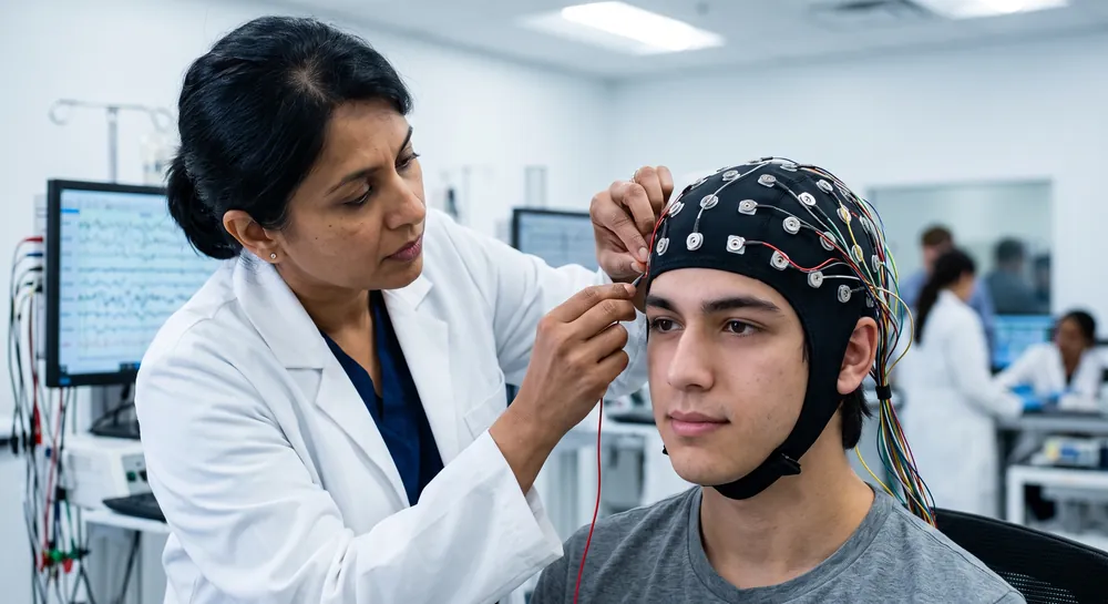
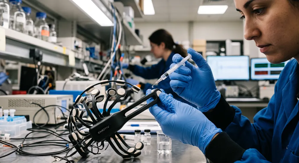
Research-grade electrophysiology, specifically quantitative EEG (qEEG), is being actively explored as a method to non-invasively measure cortical dysfunction and subclinical cognitive decline in ALS.
These tools allow researchers to investigate cortical hyperexcitability, a physiological state where neurons become overly sensitive and fire excessively, which is believed to be a central component of the disease's pathophysiology.
In studies of bulbar-onset ALS, qEEG provides a high-resolution view of how electrical signaling patterns shift as motor neurons in the brainstem and cortex deteriorate. By identifying unique electrical "signatures" of disease activity, scientists aim to refine prognostic models that currently rely on clinical measurements like functional rating scales and respiratory tests.
This neurophysiological data is crucial for identifying patients who may experience cognitive symptoms alongside physical decline, allowing for more personalized care planning. It is important to emphasize that while electrophysiological biomarkers show promise for tracking disease mechanisms and potential future prognostic use, they are currently utilized as research tools and are not yet established standards of care for clinical diagnosis or prediction.
What Future Research Directions Are Focused on the Mechanisms of Bulbar Degeneration?
Current research initiatives are pursuing multiple complementary approaches to understand and target the specific vulnerabilities of brainstem motor circuits in ALS. These efforts range from basic mechanistic studies to translational research aimed at developing bulbar-specific therapeutic interventions.
Cellular and molecular research focuses on identifying the factors that render brainstem motor neurons particularly susceptible to ALS pathology.
Meanwhile, genetic research continues to uncover new associations between specific mutations and bulbar-onset disease. Whole genome sequencing studies of large patient cohorts are identifying rare variants that may contribute to bulbar vulnerability. These discoveries could lead to genetic testing strategies that improve prognostic accuracy and guide treatment decisions.
Brain health research increasingly recognizes the importance of understanding regional vulnerability patterns in neurodegenerative diseases. Advanced neuroimaging techniques, including qEEG, diffusion tensor imaging, and functional connectivity analysis, are mapping the progression of pathology from bulbar onset sites to other brain regions.
References
Eisen, A., Vucic, S., & Mitsumoto, H. (2024). History of ALS and the competing theories on pathogenesis: IFCN handbook chapter. Clinical neurophysiology practice, 9, 1-12. https://doi.org/10.1016/j.cnp.2023.11.004
Wang, X., Hu, Y., & Xu, R. (2024). The pathogenic mechanism of TAR DNA-binding protein 43 (TDP-43) in amyotrophic lateral sclerosis. Neural regeneration research, 19(4), 800–806. https://doi.org/10.4103/1673-5374.382233
Yang, Q., Jiao, B., & Shen, L. (2020). The development of C9orf72-related amyotrophic lateral sclerosis and frontotemporal dementia disorders. Frontiers in genetics, 11, 562758. https://doi.org/10.3389/fgene.2020.562758
Dukic, S., McMackin, R., Buxo, T., Fasano, A., Chipika, R., Pinto‐Grau, M., ... & Nasseroleslami, B. (2019). Patterned functional network disruption in amyotrophic lateral sclerosis. Human Brain Mapping, 40(16), 4827-4842. https://doi.org/10.1002/hbm.24740
Frequently Asked Questions
What makes bulbar-onset ALS progress more rapidly than limb-onset?
Bulbar-onset ALS involves brainstem motor nuclei that are compactly arranged and have exceptionally high metabolic demands, making them intrinsically more vulnerable to degeneration. The bilateral redundancy of corticobulbar pathways initially masks early damage, but once functional thresholds are crossed, decline accelerates sharply.
Which cranial nerves are primarily affected in bulbar ALS?
The trigeminal motor nucleus controls chewing, and facial nucleus weakness impairs lip seal during speech and swallowing. The glossopharyngeal and vagus nuclei coordinate swallowing and voice, while hypoglossal nucleus damage causes tongue atrophy and fasciculations; accessory nucleus involvement contributes to head drop and neck weakness.
How do C9orf72 gene expansions relate to bulbar-onset ALS?
C9orf72 repeat expansions are the most common genetic cause of ALS and strongly associated with bulbar symptom onset. The mutation produces toxic dipeptide repeat proteins and RNA foci that disproportionately stress brainstem motor neurons, leading to faster disease progression.
What is the difference in TDP-43 pathology between bulbar and limb-onset ALS?
Bulbar-onset cases show more extensive and earlier accumulation of TDP-43 protein aggregates within brainstem motor nuclei and respiratory control centers. Different conformational strains of TDP-43 may preferentially target brainstem neurons, contributing to the aggressive disease course.
What is the clinical link between bulbar ALS and frontotemporal dementia (FTD)?
The two conditions share genetic drivers such as C9orf72 expansions and overlapping brain atrophy in frontal and temporal language regions. Consequently, cognitive impairment and behavioral changes occur at higher rates in bulbar-onset ALS, reflecting a spectrum of shared neurodegeneration.
Why does age at onset strongly predict prognosis in bulbar-onset ALS?
Older patients have less physiological reserve and pre-existing age-related motor neuron loss in the brainstem, so ALS pathology more quickly surpasses functional thresholds. This age-based vulnerability leads to faster declines and shorter survival, even after accounting for other factors.
How is the rate of speech decline used as a prognostic marker?
A declining speaking rate measured in syllables per second during standardized tasks reflects progressive motor control loss and predicts future functional decline. Patients who lose intelligible speech early, typically within a year, tend to have more aggressive disease with earlier respiratory involvement.
Emotiv is a neurotechnology leader helping advance neuroscience research through accessible EEG and brain data tools.
Medical Disclaimer: The information provided on this website is for educational and informational purposes only and is not intended as medical or health advice. This content may contain errors and should not be relied upon to make life-altering health, medical, or lifestyle choices. Always seek the advice of your physician or other qualified health provider with any questions you may have regarding a medical condition or treatment.
Christian Burgos