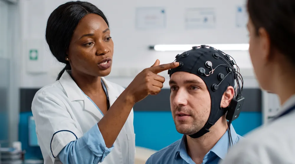
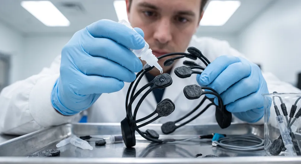
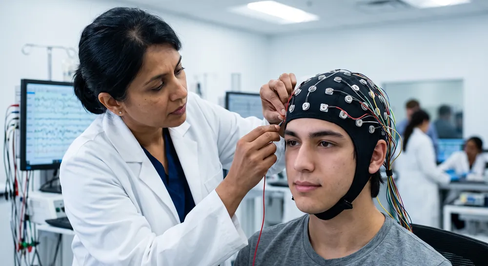
The brain’s theta rhythms have captured scientific interest for their apparent role in linking memory, performance, and emotional regulation. This article examines the current evidence for theta-band activity in electroencephalography (EEG), specifically its potential benefits in memory consolidation, executive function, musical performance, complex skill learning, and mental health.
The Role of Theta Rhythms in REM Sleep
One of the most striking theta-related findings comes from sleep research. A nap study explored how rapid eye movement (REM) sleep and its unique electrophysiology help the brain consolidate emotional memories.
Participants who napped after viewing emotionally charged and neutral images showed a selective benefit for emotional material. The No-Nap control group did not show this consolidation advantage for either memory type.
Crucially, the degree of emotional memory facilitation within the Nap group was significantly correlated with both the amount of REM sleep obtained and the power of right-dominant prefrontal theta oscillations during REM.
This pattern points to a specific role for theta activity, not in memory generally, but in the processing of emotionally salient experiences. Theta is suggested to help tag memories with affective weight, allowing them to be reinforced during sleep. The right prefrontal cortex, a region involved in emotional regulation and autobiographical memory, appears to be a key hub for this consolidation.
The findings are compelling because they link a measurable brain rhythm to a behavioral outcome in a controlled setting. However, the evidence is purely correlational and does not prove that increasing theta during sleep would strengthen emotional memory processing, nor does it demonstrate a clinical effect on mood disorders.
It does suggest that theta rhythms during REM sleep could be a biomarker for healthy emotional memory integration, and that disruptions in this process might contribute to the intrusive memories seen in anxiety or post-traumatic stress. These are translational implications worth exploring, but they remain hypotheses.
Can Theta Neurofeedback Enhance Working Memory?
Theta activity also appears in EEG protocols designed to enhance cognition while awake. A review tutorial on neurofeedback describes a training protocol that targets frontal-midline theta–a specific theta rhythm generated in the anterior cingulate and prefrontal regions–to improve executive functions such as working memory and attentional control. The protocol involved teaching individuals to voluntarily up-regulate this theta activity through real-time EEG feedback.
The review positions this as a promising intervention, illustrating the step-by-step setup of a neurofeedback study and the theoretical mechanisms by which frontal-midline theta might support cognitive control. Theta oscillations in this frequency range are thought to coordinate communication between distant brain networks, particularly during tasks that require sustained attention and mental effort. By learning to increase this signal, participants might strengthen the neural circuits underlying executive function.
Yet the evidence remains preliminary. The tutorial presents a rationale and a protocol, not a body of controlled trials.
Theta Neurofeedback for Musical Performance
Theta’s influence on real-world performance has been tested in a series of experiments with conservatoire students. In a pilot study, students received neurofeedback training aimed at enhancing attention and relaxation. The training focused on learning to progressively raise the amplitude of theta (5–8 Hz) relative to alpha (8–11 Hz).
Expert ratings of live musical performances, conducted under stressful conditions, showed significant improvements. These gains were highly correlated with the ability to increase the theta-to-alpha ratio during training.
Moreover, the alpha/theta training group displayed a clear performance enhancement, while other neurofeedback protocols and the Alexander technique did not produce comparable benefits. This suggests that the specific modulation of slow-wave activity, particularly the shift toward theta over alpha, played a causal role in the observed improvements.
The study used an ecologically valid measure: real-life music performance assessed by blind raters. This is a strength, as it moves beyond laboratory tasks. The improvements likely reflect a combination of reduced performance anxiety and enhanced automaticity, both of which are associated with a theta-dominant state.
Theta Power and Skill Acquisition in Surgery
A randomized controlled trial with trainee ophthalmic microsurgeons provides a more nuanced picture of theta’s role in skill acquisition. Participants were assigned to either Sensory Motor Rhythm-Theta (SMR) neurofeedback or Alpha-Theta (AT) neurofeedback, with a wait-list control group.
SMR training, which reinforces a low-beta rhythm while suppressing theta, led to significant improvements in surgical technique and speed. Expert ratings of overall technique and suture task performance improved substantially, with effect sizes of 0.6 and 0.9 respectively. Meanwhile, time on task decreased by 26%.
Importantly, reduced surgical time was strongly associated with the EEG changes specific to SMR training, particularly a continued reduction of theta (4–7 Hz) power.
Based on these results the authors suggested that in the SMR group, lowering theta power was linked to faster, more efficient performance. Meanwhile, the Alpha-Theta group, which aimed to increase the theta-to-alpha ratio, showed only marginal improvements in technique and overall time. However, within that group, successful within-session elevation of the theta-alpha ratio did correlate positively with better overall technique (r \= 0.64). So theta’s role depended on the training protocol and the specific outcome.
Reducing theta appeared beneficial for speed and technical precision, while increasing the theta-alpha ratio might support a different aspect of performance, perhaps a more relaxed, fluid execution style.
Can Theta Rhythms Benefit Mental Health?
The emotional memory consolidation findings from the REM sleep study carry plausible implications for mental health.
If theta activity during REM is crucial for processing emotional memories, then abnormalities in this process could contribute to mood disorders. The study explicitly mentions translational implications for affective psychiatric conditions, but it did not test any clinical outcomes. Theta power during REM could serve as a biomarker for adaptive emotional processing, but no direct therapeutic benefit has been demonstrated.
Similarly, the surgical training study showed that anxiety decreased after SMR neurofeedback, but this was in healthy individuals and the protocol involved reducing theta power. Therefore, this cannot be used to argue that increasing theta directly alleviates anxiety.
The mental health benefits of theta wave activity remain an active area of investigation, but the current evidence is sparse and insufficient to support clinical recommendations. Theta’s connection to emotional regulation is logical, given its role in limbic-cortical communication, but the leap from correlation to treatment is still unsupported by rigorous data.
Why Theta Rhythms Depend on Context, Task, and Brain Region
The studies reviewed here associate theta-band EEG activity with specific, limited benefits: emotional memory consolidation during REM sleep, enhanced musical performance under stress, and, in certain contexts, improved executive function and complex skill learning. The evidence is mixed and highly conditional.
Theta’s role varies depending on whether it is being increased or decreased, the brain region involved, and the nature of the task. No study in this set demonstrates that theta waves universally improve memory, creativity, or mental health. The findings are promising but preliminary, and they should be interpreted with caution.
As research progresses, the challenge will be to specify exactly when and how theta modulation can be harnessed safely and effectively.
References
Nishida, M., Pearsall, J., Buckner, R. L., & Walker, M. P. (2009). REM sleep, prefrontal theta, and the consolidation of human emotional memory. Cerebral cortex, 19(5), 1158-1166. https://doi.org/10.1093/cercor/bhn155
Enriquez-Geppert, S., Huster, R. J., & Herrmann, C. S. (2017). EEG-neurofeedback as a tool to modulate cognition and behavior: a review tutorial. Frontiers in human neuroscience, 11, 51. https://doi.org/10.3389/fnhum.2017.00051
Egner, T., & Gruzelier, J. H. (2003). Ecological validity of neurofeedback: Modulation of slow wave EEG enhances musical performance. Neuroreport, 14(9), 1221-1224.
Ros, T., Moseley, M. J., Bloom, P. A., Benjamin, L., Parkinson, L. A., & Gruzelier, J. H. (2009). Optimizing microsurgical skills with EEG neurofeedback. BMC neuroscience, 10(1), 87. https://doi.org/10.1186/1471-2202-10-87
Frequently Asked Questions
Does theta wave activity improve memory?
Theta activity is specifically linked to emotional memory consolidation during REM sleep, where right-dominant prefrontal theta power correlates with better recall of emotionally charged images. This association is correlational and does not prove that increasing theta would enhance memory in general.
Can theta neurofeedback enhance executive function?
A neurofeedback protocol targeting frontal-midline theta aims to improve working memory and attentional control by teaching individuals to up-regulate this rhythm. However, the evidence is preliminary, with no large-scale randomized trials confirming reliable real-world improvements.
Is reducing theta beneficial for skill learning?
In a surgical training study, SMR neurofeedback that reduced theta power improved surgical technique and speed, with lower theta linked to faster performance. This shows that decreasing theta can be beneficial for tasks requiring precision and efficiency, contrary to a simple "more theta is better" view.
What is the role of theta in emotional memory consolidation?
During REM sleep, theta power in the right prefrontal cortex correlates with the selective consolidation of emotional memories, helping tag them with affective weight. This process may be a biomarker for adaptive emotional processing, but disruptions remain a hypothesis for mood disorders.
Emotiv is a neurotechnology leader helping advance neuroscience research through accessible EEG and brain data tools.
Medical Disclaimer: The information provided on this website is for educational and informational purposes only and is not intended as medical or health advice. This content may contain errors and should not be relied upon to make life-altering health, medical, or lifestyle choices. Always seek the advice of your physician or other qualified health provider with any questions you may have regarding a medical condition or treatment.
Christian Burgos