There is a persistent problem embedded in how EEG is recorded, the voltage detected at any single electrode is not a clean readout of the brain tissue directly beneath it. It is a mixture, shaped by tissue layers, electrode placement, and an arbitrary reference point chosen by the person running the recording.
The Laplacian montage was developed specifically to address this mixture problem. Rather than reporting raw voltage, it transforms the scalp signal into an estimate of local current source density, a measure that is not tied to any external reference and that correlates more directly with the electrical activity happening in the cortex right underneath the sensor.
The sections below walk through why this transformation is necessary, how it is mathematically derived, and what the supporting research shows about its practical advantages.
What is a Laplacian Montage in EEG?
Clinical electroencephalography relies on the arrangement of scalp sensors to visualize neural activity patterns accurately. Traditional electrode montages record potentials relative to a specific reference, which can sometimes conflate signal clarity across larger surface areas. The laplacian montage EEG offers a distinct analytical alternative by focusing on local differences rather than global potentials.
Understanding the Basics of EEG Laplacian Montage
The EEG signal essentially reflects the collective electrical activity of pyramidal neurons beneath the scalp. When an electrode captures a potential, it inevitably includes contributions from distant brain sources due to the volume conduction properties of the skull and scalp.
The process of extracting these subtle rhythms requires clear methodology, often involving the grounded principles of neuroscience to ensure that the waveforms analyzed correspond to discrete localized brain regions.
Why Scalp EEG Signals Are Hard to Interpret Accurately
The brain's electrical signals do not travel in a straight line to the electrode. They pass through cerebrospinal fluid, skull bone, and scalp tissue before they can be measured, and each of these layers conducts electricity differently.
The skull, in particular, behaves like a spatial low-pass filter as it smooths and spreads the signal, blurring activity that may be quite localized in the cortex into a broad, diffuse pattern by the time it reaches the scalp.
Research (Srinivasan et al.) modeling the head as four concentric spherical layers (brain, cerebrospinal fluid, skull, and scalp) has shown that this spreading is strong enough to make electrodes as far as 10 to 12 centimeters apart appear artificially correlated, even when the underlying neural sources are entirely unrelated. This creates a real risk of interpreting correlated scalp readings as evidence of coordinated brain activity, when the correlation may be nothing more than an artifact of how electricity diffuses through tissue.
A second distortion comes from the reference electrode itself. Conventional EEG montages report voltage as a difference between an active electrode and a reference point, but that reference is never electrically silent.
Simulation studies and empirical recordings (Nunez et al.) have demonstrated that the choice of reference can shift the apparent timing of brain events, meaning the latency of an evoked response recorded with one reference scheme may not match the latency recorded with another. This is a subtle but consequential problem, since much of EEG's clinical and research value depends on precise timing.
A third source of contamination is muscular, not neural. Central and pericentral scalp sites, the electrodes positioned over the top and sides of the head, sit close to scalp and jaw musculature. Electrical activity from these muscles readily leaks into the recording, particularly at higher frequencies, and conventional referencing schemes do little to separate muscle-generated signal from brain-generated signal.
Together, volume conduction, reference dependence, and muscle contamination form three compounding reasons why raw scalp potentials give an imprecise picture of what the cortex is actually doing.
Problem | Description |
|---|---|
Volume conduction | Skull blurs and spreads signals |
Reference electrode dependency | Reference choice distorts event timing |
Muscle contamination | EMG leaks into central electrodes |
What Is the Surface Laplacian and How Does It Work
The surface Laplacian addresses these problems by changing what is being measured. Instead of recording voltage directly, it calculates the second spatial derivative of the voltage field across the scalp, essentially asking how sharply the potential is curving at each point on the head rather than what its absolute value is.
This curvature measurement is proportional to the radial current flowing into and out of the scalp at that location, which makes it a physical estimate of local current source density rather than a raw electrical reading influenced by distant activity.
Because differentiation is a mathematical operation that removes constant offsets, this approach has a built-in advantage: any voltage that is uniformly added to every electrode, which is exactly what happens when a shared reference electrode is used, cancels out during the calculation.
The result is a signal that no longer depends on the placement of a reference at all. This is why the Laplacian is often described as reference-free.
The Laplacian also functions as what researchers describe as a spatial bandpass filter. It suppresses very broad, diffuse patterns of voltage change (the kind produced by volume conduction spreading across large regions of the scalp) while also attenuating extremely sharp, focal noise.
What remains is a moderate-scale estimate of activity that appears to correspond well with how electrical currents from the cortex actually propagate through the layers of the human head. In effect, the transformation is tuned to the physical scale at which neocortical sources genuinely influence the scalp, filtering out both the too-broad and the too-narrow.
Reference Electrode Standardization Technique (REST)
Before applying the Laplacian transformation, the choice of the primary physical reference often influences the initial recording quality.
Many clinics utilize the Reference Electrode Standardization Technique (REST), which mathematically transforms the raw EEG data to an approximate reference-independent distribution. This ensures that the subsequent computation is not skewed by the specific electrical site chosen for the initial recording, which is critical for objective clinical assessment.
How the Spline-Laplacian Is Computed in Practice
Calculating a second derivative from a finite set of scattered electrode readings is not straightforward, since electrodes only sample the scalp at discrete points rather than continuously.
The spline-Laplacian method solves this by fitting a smooth, flexible mathematical surface, modeled as a sphere or a more anatomically realistic ellipsoid, through the actual electrode positions. Once this continuous surface is defined, the derivative can be computed directly from it, producing a Laplacian estimate at every electrode location based on the values recorded at its surrounding neighbors.
This method was originally derived for spherical head models and was later extended mathematically to ellipsoidal surfaces, which better approximate the actual shape of a human head. Both derivations have been shown to remain stable even when there are inaccuracies in head geometry or uncertainty about the resistivity of different tissue layers, factors that are essentially unavoidable in real clinical or research recording sessions.
This robustness means the spline-Laplacian does not require a perfect anatomical model of an individual's head to produce a useful and stable result.
There is one practical requirement that determines how much benefit the method delivers: electrode density. Research by Nunez et al. comparing spline-Laplacian performance across different electrode layouts found dramatic improvement in spatial resolution specifically when the average spacing between neighboring sensors is smaller than approximately 3 centimeters.
Below this spacing, the derivative can be estimated with enough precision to sharpen the underlying signal substantially. Sparse electrode arrays, by contrast, do not sample the scalp finely enough to support an accurate second-derivative calculation, limiting how much the transformation can improve on raw potentials.
Calculating the Laplacian Potential
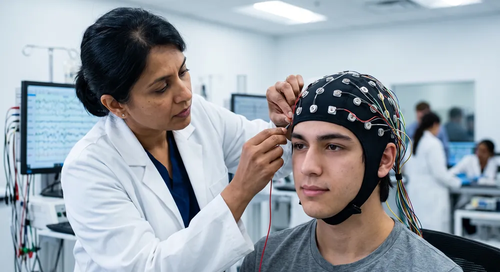
To compute the potential, a software system assesses the center sensor against a weighted average of its immediate neighbors in a radial pattern. This creates a virtual map of current density, which is often easier to interpret during diagnostics.
The core of the mathematical sequence for this computation is detailed below:
Step | Action | Purpose |
|---|---|---|
1 | Electrode Selection | Choose the central point of analysis. |
2 | Spatial Weighting | Apply values to neighboring scalp sensors. |
3 | Gradient Computation | Subtract the local average from the center. |
The following criteria help determine if the configuration is optimized for clear results:
Inter-electrode distance must remain uniform where possible.
Signal quality at all surrounding neighbors must be comparable.
The configuration should maintain symmetry around the zone of interest.
Once these criteria are met, the resulting data effectively highlight the focal source of brain activity, showing reduced interference from far-field patterns.
Advantages of Using a Laplacian Montage
Spatial filtering provides several distinct benefits for researchers aiming to isolate specific cortical generators. By reducing reliance on a single reference point, the technique fosters more reliable results across different experimental conditions.
Improved Spatial Resolution With the Laplacian Transform
The central practical claim behind the Laplacian montage is that it sharpens the spatial picture of brain activity considerably compared with unprocessed scalp voltage.
The work by Nunez et al. using spline-based derivations on spherical and ellipsoidal surfaces reported spatial resolution improvements of at least a factor of three over conventional recordings. This improvement held up across computer simulations, evoked potential data, spontaneous resting EEG, and recordings of epileptic spikes, suggesting it is not limited to one narrow type of brain signal.
A separate analysis by Law et al. reinforced this finding by showing that the improvement in resolution is largely independent of the specific assumptions made about the source of the signal or the geometric model used to represent the head. This is an important distinction.
Many EEG source-localization techniques require researchers to make prior assumptions about where in the brain a signal is likely coming from. The spline-Laplacian achieves its resolution gains without depending heavily on those assumptions, which makes it more broadly applicable across different types of studies and patient populations, provided electrode density is sufficient.
Removing Reference Electrode Distortion
Because the Laplacian calculation mathematically cancels out any constant value added across all electrodes, it eliminates the influence of the reference electrode by construction rather than by choosing a supposedly neutral reference site.
The comparative work by Nunez et al. examining potential data directly demonstrated that raw scalp potentials, still tied to whatever reference was selected, can distort the apparent shape and timing of an event-related brain response. The current source density estimate produced by the Laplacian transform, by contrast, was shown to provide a more accurate spatio-temporal description of the same underlying event.
In practical terms, this means two labs using different reference electrodes on the same subject could report meaningfully different-looking waveforms from raw potentials, while their Laplacian-transformed data would converge on a more consistent representation of the underlying cortical activity.
Reducing Artificial Coherence From Volume Conduction
Coherence, a statistical measure of how similarly two signals fluctuate over time, is commonly used in EEG research to infer whether two brain regions are communicating or working together. The problem is that volume conduction alone, with no actual coordinated neural activity involved, can generate high coherence values between nearby electrodes simply because the underlying voltage has spread across the scalp.
Using an analytic model of the head's layered conductivity, researchers in Srinivasan’s group demonstrated that this volume-conduction effect can produce artificial correlation between electrodes up to 10 to 12 centimeters apart. Applying the surface Laplacian to the same data reduced this artificial coherence substantially, because its spatial bandpass properties filter out exactly the kind of broad, diffuse spreading that produces false correlation.
This does not mean raw potential coherence should be discarded outright. The same research emphasized that raw scalp coherence and Laplacian-derived coherence are sensitive to different spatial bandwidths of cortical activity, meaning each captures a somewhat different slice of neocortical dynamics.
Rather than replacing one measure with the other, the recommendation is to examine both in parallel, since together they offer a more complete picture than either alone.
Temporal Accuracy: Why Latency Estimates Improve
EEG's reputation rests heavily on its speed, its ability to track brain activity on a millisecond timescale. That reputation is somewhat overstated when applied to raw scalp potentials.
The simulation work aforementioned has shown that volume conduction and reference electrode choice do not just distort where a signal appears to originate, they also distort when it appears to happen. Scalp potentials can mis-estimate the latency of genuine brain events because the smearing effect of tissue conduction and the influence of the reference blend signals from different time points and different sources together.
The same body of work found that current source density estimates generated through the surface Laplacian avoid much of this distortion, offering what the researchers described as a much richer and much more accurate view of the spatio-temporal dynamics of brain activity. This finding was replicated across two simulation studies and two empirical datasets, giving it a fairly consistent evidence base.
The practical implication is that researchers studying the precise timing of cognitive or clinical events, not just their spatial origin, have reason to consider Laplacian-transformed data as a more trustworthy record of when things are actually happening in the brain.
Muscle Artifact Rejection in Central Scalp Leads
Muscle-generated electrical activity, or electromyographic contamination, is one of the more stubborn confounds in EEG recording, particularly at central scalp sites near jaw and scalp musculature.
A study by Fitzgibbon et al. designed to isolate this effect compared recordings taken from awake subjects before and after complete neuromuscular blockade, which allowed researchers to measure how much of the recorded signal in normal conditions was actually muscle rather than brain activity.
Comparing several scalp surface Laplacian estimators against left-ear reference and common average reference montages, the study found that surface Laplacian processing reduced muscle power in central and pericentral leads to less than one sixth of the brain signal above 30 hertz, a brain-to-muscle ratio greater than six.
This performance was reported as two to three times better than common average reference, one of the more widely used conventional montages. Because muscle contamination tends to concentrate in higher frequency ranges, this advantage is particularly relevant for anyone trying to study gamma-band activity, a frequency range of clinical and cognitive interest that is otherwise easily obscured by scalp and jaw muscle noise.
The researchers noted this makes the Laplacian a useful standard for detecting high-frequency activity and for studying electrophysiological correlates of disease, including conditions studied within brain disorders research, where subtle high-frequency signals may carry diagnostic weight.
Applications of Laplacian Montage EEG
Clinical assessment of epilepsy remains one of the primary applications for this spatial processing method. By identifying the exact spatial distribution of interictal discharges, neurologists can better define the seizure focus. This provides a clearer view than standard recordings, which often present with significant blurring due to surrounding cranial anatomy.
Cognitive neuroscience research also employs this approach, particularly when probing high-frequency oscillations that require precise timing and location. Studies often track these pulses across the cortical surface to observe how they travel between sensory processing hubs.
Finally, the technique is widely used in Brain-Computer Interface (BCI) development where real-time accuracy is essential for motor control. By isolating the specific mu rhythms generated in the motor cortex, the system can interpret intent more accurately.
This application demonstrates the versatility of Laplacian filters in turning raw electrical potentials into functional input for external devices.
Limitations and Interpretation Caveats
None of these advantages make the Laplacian a universal replacement for other EEG analysis approaches, and the supporting research is explicit about its boundaries.
First, the Laplacian is not a source localization technique in the sense of pinpointing an exact anatomical location for a signal. It produces an estimate of current density at a moderate spatial scale, which is a different goal than the kind of localization performed by dipole-fitting or other model-based methods.
Second, the transformation is described as insensitive to sources that originate deep within the brain, away from the cortical surface, or to sources located outside the physical boundary of the electrode array itself. If a signal comes from subcortical structures or from a region the electrode net does not cover, the Laplacian will not represent it well, regardless of how densely the surrounding electrodes are placed.
Third, the resolution gains are conditional. Substantial improvement depends on electrode spacing averaging less than roughly 3 centimeters, so a sparse or unevenly spaced array will not deliver the same benefit demonstrated in the underlying studies. Anyone applying the method to lower-density recordings should expect more modest gains.
Finally, the same spatial bandpass property that filters out volume-conduction artifact can also attenuate genuinely widespread cortical events, since very broad patterns of activity resemble the diffuse signals the filter is designed to suppress.
This is why the coherence research recommended analyzing raw potential data and Laplacian-transformed data in parallel rather than treating one as a strict upgrade over the other. Each captures a different spatial bandwidth of neocortical activity, and the most complete interpretation comes from considering both together.
Conclusion: The Laplacian as a Sharper Lens on Cortical Activity
The surface Laplacian reframes what scalp EEG is measuring. Rather than reporting a voltage that depends on an arbitrary reference and that has been blurred by the skull's filtering effect, it estimates local current source density directly from the geometry of the electrode array, using spline-based methods that have been shown to remain stable under real-world head modeling errors.
The empirical record built across these studies points to consistent, measurable advantages:
Spatial resolution improved by a factor of three or more
Artificial correlation between distant electrodes suppressed
Latency estimates that better reflect actual brain timing
Muscle contamination reduced to a fraction of what conventional referencing allows
These gains depend on adequate electrode density and come with real interpretive limits, particularly around deep or out-of-array sources and the risk of attenuating broad cortical patterns. Used alongside raw potential analysis rather than as its replacement, the Laplacian montage offers a meaningfully sharper, reference-free window into local cortical activity.
References
Srinivasan, R., Nunez, P. L., & Silberstein, R. B. (1998). Spatial filtering and neocortical dynamics: estimates of EEG coherence. IEEE transactions on Biomedical Engineering, 45(7), 814-826. https://doi.org/10.1109/10.686789
Nunez, P. L., & Pilgreen, K. L. (1991). The spline-Laplacian in clinical neurophysiology: a method to improve EEG spatial resolution. Journal of Clinical Neurophysiology, 8(4), 397-413.
Law, S. K., Nunez, P. L., & Wijesinghe, R. S. (2002). High-resolution EEG using spline generated surface Laplacians on spherical and ellipsoidal surfaces. IEEE transactions on Biomedical engineering, 40(2), 145-153. https://doi.org/10.1109/10.212068
Fitzgibbon, S. P., Lewis, T. W., Powers, D. M., Whitham, E. W., Willoughby, J. O., & Pope, K. J. (2012). Surface laplacian of central scalp electrical signals is insensitive to muscle contamination. IEEE Transactions on Biomedical Engineering, 60(1), 4-9. https://doi.org/10.1109/TBME.2012.2195662
Frequently Asked Questions
What is the surface Laplacian in EEG analysis?
The surface Laplacian estimates the second spatial derivative of the scalp voltage field, which corresponds to the radial current flowing into and out of the scalp. This transforms the recording into a measure of local current source density rather than raw voltage, making it largely independent of the reference electrode.
How does the Laplacian montage eliminate the reference electrode problem?
The Laplacian calculation mathematically cancels out any constant voltage that is added uniformly to all electrodes, which is exactly what a shared reference does. Because of this built-in cancellation, the resulting signal no longer depends on where the reference electrode was placed.
What role does the Laplacian play in reducing volume conduction artifacts?
The Laplacian acts as a spatial bandpass filter that suppresses broad, diffuse voltage patterns caused by volume conduction through the skull and scalp. This filtering reduces the artificial coherence between distant electrodes that would otherwise be misinterpreted as coordinated brain activity.
How does the Laplacian improve the timing accuracy of EEG signals?
Volume conduction and reference choice can smear the timing of brain events in raw scalp potentials. The Laplacian’s current source density estimate reduces this smearing, providing a more accurate representation of when cortical activity actually occurs.
Why is a high electrode density important for the spline-Laplacian method?
The spline-Laplacian computes a second derivative from a discrete set of electrode readings, so the scalp must be sampled finely enough to capture the voltage curvature. When average sensor spacing is sufficiently small, the derivative can be estimated with precision, yielding substantial gains in spatial resolution.
Can the Laplacian help reduce muscle artifact in EEG?
Yes, surface Laplacian processing significantly reduces muscle-generated electrical noise, especially at central scalp sites near jaw and scalp muscles. This results in a much higher ratio of brain signal to muscle contamination, particularly in higher frequency ranges like gamma.
What are the main limitations of the Laplacian montage?
The Laplacian does not locate deep brain sources or signals outside the electrode array, and it can attenuate genuinely widespread cortical activity because its filter suppresses broad patterns. It is best used alongside raw potential analysis, as each captures a different spatial scale of brain activity.
How does the Laplacian montage differ from a bipolar montage?
A bipolar montage compares two distinct electrodes to show voltage differences, while the Laplacian montage uses a mathematical second derivative based on a center electrode and its immediate neighbors to estimate local current density across a surface.
Does the technique require a specific number of electrodes?
Yes, the efficacy of the montage scales with channel count, as the calculation depends on the spatial density of the sensor array and the relative accuracy of the neighbor grid layout.
Can Laplacian montages be used with standard 10-20 system layouts?
While mathematically possible with limited electrodes if using specialized interpolation, standard 10-20 systems may lack the density required for a highly reliable or detailed spatial interpretation.
Can the Laplacian montage detect deep brain structures?
Because the montage acts as a spatial high-pass filter, it is designed to emphasize superficial cortical activity and is generally less sensitive to deep subcortical sources compared to potential-based displays.
Emotiv is a neurotechnology leader helping advance neuroscience research through accessible EEG and brain data tools.
Medical Disclaimer: The information provided on this website is for educational and informational purposes only and is not intended as medical or health advice. This content may contain errors and should not be relied upon to make life-altering health, medical, or lifestyle choices. Always seek the advice of your physician or other qualified health provider with any questions you may have regarding a medical condition or treatment.
Christian Burgos