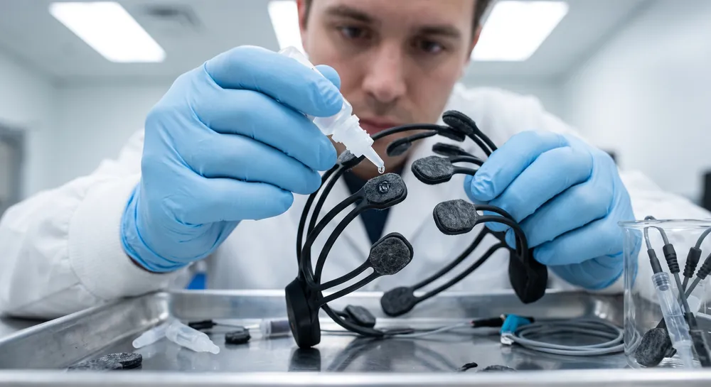
Every electroencephalogram trace on a readout is the product of a choice. That choice determines whether a spike of electrical activity on the page reflects a single point on the scalp or the relationship between two points.
Bipolar recording is one of the two dominant ways to make that choice, and understanding how it works requires stepping back into basic circuit logic before returning to the EEG lab. The method is old, taught in nearly every clinical neurophysiology course, and still forms the backbone of automated detection systems built to catch seizures and spikes in real time.
What Is Bipolar Montage in EEG?
A standard EEG electrode captures a voltage relative to some reference point, often a distant or averaged location on the scalp.
A bipolar channel does something different. It records the voltage difference between two adjacent electrodes, for example the pairing of Fp1 and F7, and displays that difference as a single trace. The math behind each channel is simple: take the instantaneous voltage at electrode A, subtract the instantaneous voltage at electrode B, and plot the result.
This arrangement appears directly in applied research on automated seizure detection. In a 2013 physiology-based detection system built for multichannel EEG, Shen et al. analyzed both unipolar and bipolar signals side by side, treating the bipolar format as a legitimate and necessary input alongside the single-point measurements.
Furthermore, a separate classification model built to distinguish focal from generalized epilepsy went further, constructing its entire feature set around a longitudinal bipolar montage, a specific chain of adjacent electrode pairs running from front to back across the scalp. In that 2022 study by Najafi et al., the bipolar format was not an alternative option considered among several. It was the foundation the whole model was built on.
The practical reason bipolar recording persists across decades of clinical practice and modern machine learning pipelines alike comes down to what happens mathematically when you subtract two signals that share a common source of interference. That mathematical behavior is where the real value of the montage begins.
Electrode Placement and Referencing
Proper electrode placement is essential to ensure that the detected electrical activity is accurately representative of regional brain function. Clinicians and researchers typically adhere to established protocols to maintain symmetry and consistency across varied patient populations. The signal processing involves specific configurations, as outlined below, to isolate neurological signals.
Configuration Type | Channel Input 1 | Channel Input 2 |
|---|---|---|
Longitudinal Bipolar | Frontal Electrode | Central Electrode |
Transverse Bipolar | Temporal Electrode | Temporal Electrode |
Sequential Trace | Active Point A | Active Point B |
By comparing adjacent sites, the electrodes provide a clear view of local fluctuations. This setup prevents the common mode rejection of signals that occurs in other referencing methods, allowing for sharper focal signal spikes during interpretation.
Interpreting the Bipolar EEG Montage
Interpreting the resulting data requires an understanding of the phase reversals and voltage gradients across the grid.
When a potential difference occurs at a specific electrode contact, the signal indicates activity in a spatially constrained cortical area. This allows for precise anatomical localization, provided the signal generators are aligned with the chain of electrodes being recorded.
The Physics of Sequential Subtraction
Any electrical signal picked up equally by two neighboring electrodes will vanish when one is subtracted from the other. This is the basic logic of a differential measurement, and it explains why bipolar recordings are traditionally described as noise-resistant.
Consider a source of interference that is not coming from the brain directly beneath the electrodes, but from somewhere far away: muscle tension in the jaw, electrical hum from nearby equipment, or a distant brain region whose electrical field spreads broadly across the scalp.
If that “far-field” signal reaches two adjacent electrodes with roughly equal strength, subtracting one from the other cancels it out. Engineers call this common-mode rejection, and it is a foundational principle in the design of biopotential amplifiers used across electroencephalogram recording generally, not just in EEG.
It is worth being precise about what is being claimed here and what is not. This noise-cancellation property is a long-standing, widely accepted inference from signal theory, taught as a near-universal principle in clinical neurophysiology training.
Converting Spatial Voltage Gradients Into Deflections
Once far-field noise is set aside, what remains in a bipolar channel is a measurement of something specific: how much the voltage changes across the short distance between two electrodes. This is often described as a spatial gradient, meaning the trace reflects a rate of change in the electrical field along the direction of the electrode chain, rather than an absolute reading at one location.
The direction of the deflection follows a simple rule. If the first electrode in a pair is more positive than the second, the trace deflects one way, conventionally upward in most clinical recording conventions. If the polarity flips, so does the direction of the trace.
The size of that deflection is not arbitrary either. A steeper change in voltage over that short inter-electrode distance produces a larger deflection, while a shallow, gradual change produces a smaller one.
This becomes useful when measuring activity that moves across the cortex over time. As a wave of neuronal depolarization spreads across a region of tissue, the point of maximum voltage shifts along with it.
In a chain of bipolar electrodes running across that region, this produces a predictable, sequential pattern of upward and downward deflections moving from one channel to the next, effectively tracing the movement of the electrical wavefront across adjacent channels.
Phase Reversal: The Localizing Signature
Phase reversal is arguably the single most useful pattern that bipolar recording makes visible. It occurs when a focal source of electrical activity in the cortex lies directly beneath an electrode that is shared between two adjacent bipolar channels.
Picture three electrodes in a row, and two bipolar channels built from them: the first pairing electrodes one and two, the second pairing electrodes two and three.
If the true electrical source sits underneath electrode two, the two channels will show deflections that point in opposite directions at the exact same moment. One trace swings up while the other swings down, even though both are reacting to the same underlying event.
This opposite-polarity pattern is what researchers call a phase reversal, and its diagnostic value comes from what it points to. The electrode common to both reversing channels, electrode two in this example, marks the location of the steepest voltage gradient on the scalp, and by inference, the location closest to the underlying neuronal generator producing the abnormal activity.
This is the mechanism that allows a trained reader to look at a page of bipolar traces and identify not just that a seizure or spike occurred, but roughly where on the scalp it originated.
The clinical weight given to this pattern is reflected directly in the design of automated detection tools. The physiology-based multichannel detection system aforementioned explicitly incorporated phase reversals and the concept of potential fields, the way voltage is distributed across the scalp during a bipolar recording, as core features fed into its classification algorithm. That design choice reflects how central phase reversal is considered within clinical neurophysiology as a category of evidence.
Applications of Bipolar Montage EEG
Diagnosing Neurological Conditions
Bipolar EEG montages are frequently employed when clinicians need to localize specific areas of abnormal neuronal activity, particularly in cases where focal epilepsy is suspected. By observing the spatial distribution of voltage changes, practitioners identify the relative epicenter of a discharge.
This diagnostic capability is essential for correlating electrical findings with specific clinical observations during evaluations.
Transverse Bipolar Montage EEG in Seizure Monitoring
This technique allows for the rapid identification of asymmetries between the hemispheres of the brain. When electrodes are linked across the scalp, any deviation from established waveforms becomes apparent immediately.
This method is particularly useful in environments where continuous observation is necessary to assess the duration and nature of seizure events without interference from shared reference points.
Research Using EEG Longitudinal Bipolar Montage
Researchers utilize these longitudinal chains to study the spread of electrical activity across the primary functional lobes of the brain. The consistent spacing between electrodes allows for mathematical modeling of wave propagation over time.
Recent studies into how conscious breathing impacts brainwaves involve analyzing these propagation patterns to determine how physiological states modulate cortical excitability. To maintain accurate records, the following steps are generally performed during the study:
Prepare the scalp with conductive paste to reduce impedance.
Apply electrodes according to the standardized 10-20 spatial system.
Verify the impedance of each individual lead against accepted standards.
Calibrate the recording hardware to ensure linear signal amplification.
Advantages and Limitations of Bipolar Montages
One primary advantage of this methodology is its immunity to potential variations at a single reference electrode site, which often complicates other recording techniques. By focusing on the difference between adjacent pairs, researchers and clinicians minimize the chance of attributing a localized signal to a faulty reference point. This creates a predictable baseline that enhances the reproducibility of findings across multiple recording sessions on the same patient.
Conversely, a limitation arises when large-scale potentials are generated across broad brain regions. Because the configuration depends on local differences, an activity that affects the entire scalp equally may appear diminished or canceled out entirely. This can obscure generalized epileptiform discharges that might be better captured by a different mounting strategy, limiting its utility in specific diagnostic scenarios.
Therefore, researchers and clinicians must remain cognizant of these dynamics when selecting the appropriate array for their study. While highly efficacious for identifying localized anomalies, the configuration should be supplemented with other methods when a broad clinical assessment is required. Achieving a balanced view allows for the triangulation of findings, ensuring the most accurate assessment of the patient's neurological status.
The Future of Bipolar Montage EEG
The trajectory of clinical observations suggests a shift toward more integrated hardware that allows for real-time switching between mounting configurations.
As computational power increases, the ability to reformat raw data into various display modes will provide greater flexibility in clinical settings. This evolution will likely reduce the time required for setup and improve the diagnostic yield in complex cases where activity patterns are not immediately obvious.
Advancements in electrode design and signal filtering will also play a role in reducing the noise floor of these recordings, leading to higher resolution in the bipolar signal display. By mitigating technical artifacts, the sensitivity to subtle cortical changes can be improved. This development will assist practitioners in diagnosing early-stage conditions where the signal-to-noise ratio is historically a primary challenge to clinical identification.
Looking toward automated analysis, the integration of algorithmic diagnostic tools will assist in the rapid screening of long-duration recordings. While the human clinician remains central to the final interpretation, these tools will provide an initial pass that flags potential regions of interest within the bipolar chains. Such synergy represents the next step in enhancing the efficiency and utility of scalp-based neurological diagnostics in standard care environments.
Conclusion
Bipolar montage remains a cornerstone of EEG application, offering a precise method for defining localized neuronal events that might otherwise be missed. By leveraging the difference between adjacent scalp locations, it provides a stable and reliable diagnostic window that is essential for accurate neurological assessment.
As research and technology continue to evolve, the application of this technique will remain central to our continued ability to decode complex cerebral activity patterns.
References
Shen, C. P., Liu, S. T., Zhou, W. Z., Lin, F. S., Lam, A. Y., Sung, H. Y., Chen, W., Lin, J. W., Chiu, M. J., Pan, M. K., Kao, J. H., Wu, J. M., & Lai, F. (2013). A physiology-based seizure detection system for multichannel EEG. PloS one, 8(6), e65862. https://doi.org/10.1371/journal.pone.0065862
Najafi, T., Jaafar, R., Remli, R., & Wan Zaidi, W. A. (2022). A classification model of EEG signals based on RNN-LSTM for diagnosing focal and generalized epilepsy. Sensors, 22(19), 7269. https://doi.org/10.3390/s22197269
Frequently Asked Questions
What is a bipolar EEG recording?
A bipolar recording measures the voltage difference between two adjacent electrodes rather than referencing a single distant point. The trace represents the instantaneous subtraction of one electrode’s voltage from the other’s, capturing local electrical activity between that pair.
How does subtraction in bipolar recording reduce noise?
When two neighboring electrodes pick up the same far-field interference, subtracting one from the other cancels that common signal out. This differential measurement, called common-mode rejection, makes bipolar channels less sensitive to distant noise like muscle tension or electrical hum.
What is a spatial voltage gradient in bipolar EEG?
A spatial gradient is the rate at which voltage changes across the scalp over the short distance between two electrodes. Bipolar traces reflect this gradient: a steep voltage difference produces a large deflection, while a shallow difference yields a small one.
What is phase reversal and how does it localize brain activity?
Phase reversal occurs when two adjacent bipolar channels sharing a middle electrode show opposite-polarity deflections at the same moment. The electrode common to both channels marks the location of the steepest voltage gradient, pointing to the likely source of the underlying brain activity.
Why are bipolar montages used in automated seizure detection systems?
Bipolar montages provide noise-resistant signals and highlight clinically useful patterns like phase reversals and spatial gradients. Automated systems can use these features to classify abnormal brain activity with high accuracy, as demonstrated in studies that built detection models around bipolar data.
How did one study use bipolar signals to distinguish focal from generalized epilepsy?
The study decomposed bipolar channel signals using wavelet transform, extracting frequency-based features for a recurrent neural network. The model classified recordings as normal or epileptic, and further separated focal seizures from generalized ones based on statistical patterns in the bipolar montage.
What are the main limitations of the evidence presented in this article?
The two studies did not directly test the noise-cancellation or localization principles against other recording methods. Their strong results come from specific patient groups, so the findings do not prove bipolar superiority or guarantee identical performance across wider populations.
How does a bipolar montage differ from a referential montage?
A bipolar montage records the difference between two active electrodes on the scalp, whereas a referential montage records the difference between an active electrode and a single, static reference point.
Why is electrode placement critical in bipolar EEG?
Because the montage calculates differences between adjacent sites, consistent placement is necessary to ensure that the signals are spatially tied to the intended regions of the cortex.
Can bipolar EEG detect generalized brain activity?
It is less effective for generalized activity because the recording method may subtract out signals that are present at equal intensity at both chosen electrode locations.
Is the bipolar montage used alone in clinical practice?
It is rarely used in isolation; standard clinical practice usually involves reviewing EEG data in multiple varied montage configurations to get a complete picture of brain activity.
Emotiv is a neurotechnology leader helping advance neuroscience research through accessible EEG and brain data tools.
Medical Disclaimer: The information provided on this website is for educational and informational purposes only and is not intended as medical or health advice. This content may contain errors and should not be relied upon to make life-altering health, medical, or lifestyle choices. Always seek the advice of your physician or other qualified health provider with any questions you may have regarding a medical condition or treatment.
Christian Burgos